Review
. 2017 May 20;18(5):1102.
doi: 10.3390/ijms18051102.

Nanoparticles as Theranostic Vehicles in Experimental and Clinical Applications-Focus on Prostate and Breast Cancer

Affiliations
Review

Nanoparticles as Theranostic Vehicles in Experimental and Clinical Applications-Focus on Prostate and Breast Cancer

Jörgen Elgqvist. Int J Mol Sci. .

Abstract

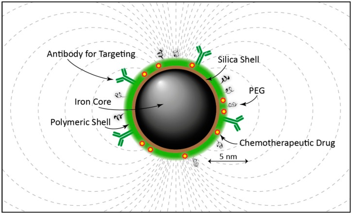
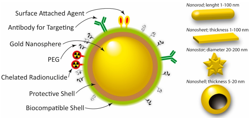
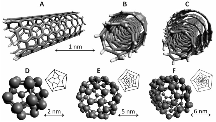
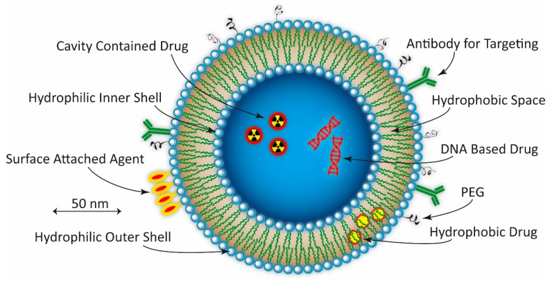
Prostate and breast cancer are the second most and most commonly diagnosed cancer in men and women worldwide, respectively. The American Cancer Society estimates that during 2016 in the USA around 430,000 individuals were diagnosed with one of these two types of cancers, and approximately 15% of them will die from the disease. In Europe, the rate of incidences and deaths are similar to those in the USA. Several different more or less successful diagnostic and therapeutic approaches have been developed and evaluated in order to tackle this issue and thereby decrease the death rates. By using nanoparticles as vehicles carrying both diagnostic and therapeutic molecular entities, individualized targeted theranostic nanomedicine has emerged as a promising option to increase the sensitivity and the specificity during diagnosis, as well as the likelihood of survival or prolonged survival after therapy. This article presents and discusses important and promising different kinds of nanoparticles, as well as imaging and therapy options, suitable for theranostic applications. The presentation of different nanoparticles and theranostic applications is quite general, but there is a special focus on prostate cancer. Some references and aspects regarding breast cancer are however also presented and discussed. Finally, the prostate cancer case is presented in more detail regarding diagnosis, staging, recurrence, metastases, and treatment options available today, followed by possible ways to move forward applying theranostics for both prostate and breast cancer based on promising experiments performed until today.

Keywords: breast cancer; nanomedicine; nanoparticles; prostate cancer; theranostics.


Conflict of interest statement

The author declares no conflict of interest.

Figures

Figure 1
Figure 1
Functionalized iron-oxide nanoparticle. Functional biocompatible polymers are grafted onto an inorganic core of magnetite (Fe3O4) or maghemite (Fe2O3) through an anchoring group, such as an amine, carboxylic acid or phosphonic acid group. The polymeric shell improves the stability of the iron oxide nanoparticles (NPs) in solution, and also allows the encapsulation of, for example, therapeutic agents. An alternative coating in the form of a fluorescent silica (SiO2) produces a type of iron oxide NPs often referred to as SCIONs, i.e., silica coated iron oxide NPs. It should be noted that the polymeric shell has to be very opaque in order not to block the fluorescent silica-based core, if used simultaneous. Most commonly, experiments with only a polymeric or a silica core have been evaluated. The custom size of an iron oxide NP is in the range of 10–20 nm, as exemplified by a 10 nm in diameter iron core in the figure. Indicated in the figure are also the magnetic field lines created by this type of NP.
Figure 2
Figure 2
Image showing an example of a nanosphere of gold (Au). Gold nanoparticles (NPs) can also be based on, for example, nano-rods, -sheets, -stars, -or nanoshells, as indicated to the right with typical dimensions. The Au-nanospheres are most often produced in the size interval of 5 to 400 nm in diameter, approximately. The characteristics of AuNPs, and therefore their application possibilities, strongly depend on size, shape, and surface functionalities. Indicated in the image are also a protective and a biocompatible shell encapsulating the Au nanosphere, enabling the attachment of different kind of targeting vectors, imaging agents, therapeutic drugs, as well as polyethylene glycol (PEG) molecules. The latter an important parameter in order to protect the NPs from the immune system, to avoid reticuloendothelial system (RES) uptake, and to minimize nonspecific binding.
Figure 3
Figure 3
Examples of carbon-based nanotubes and fullerenes. (A) Single-walled carbon nanotube (SWCNT); (B) Double-walled carbon nanotube (DWCNT); (C) Multi-walled carbon nanotube (MWCNT); (D) Fullerene based on 20 carbon atoms (20-fullerene); (E) Fullerene based on 60 carbon atoms (60-fullerene); and (F) Fullerene based on 100 carbon atoms (100-fullerene). The top size indicator applies for panels AC. The 3D-structures were created using Avogadro molecule editor. For panels AC a rod-based representation, and for panels DF a ball-based representation, was chosen for best clarity of the 3D-distribution of carbon atoms and the covalent bindings between them. For panels DF is also shown 2D-representations of the fullerene structures, as well as individual size indicators. Note, the 100-fullerene has an obloid-like structure for its global energy minima, compared to the spherical 20- and 60-fullerenes, as discussed, for example, by Yoshida et al. [225].
Figure 4
Figure 4
Schematic presentation of a spherical PEGylated liposome. Indicated are the different available spaces and surfaces that could carry different kind of targeting molecules as well as therapeutic drugs and imaging agents. In this hypothetical example, an antibody chelated to the outer surface is used for targeting. On that surface is also attached an imaging agent. Some kind of therapeutic drug is carried by the hydrophobic space between outer and inner shell. And in the core cavity, two hydrophilic drugs are situated, here exemplified by a radionuclide and a DNA-based drug such as strings of DNA, RNA, or small-interference RNA (siRNA). Unilamellar liposomes lies mostly in the size range of >150 nm, exemplified by a 200 nm liposome in the figure.
Figure 5
Figure 5
Schematic presentation of one type of spherical PEGylated dendrimer based on a central core (A), which in itself can be a dendrimer or some other type of NP (i.e., if so, a NP-cored dendrimer (NPCD)). A standard size of a dendrimer shown in A is 5–10 nm, exemplified by a 10 nm dendrimer in the figure. The surface monomers enable attachment of imaging and/or drug payloads, also able to be entrapped in the void space. The network of covalently bound interior monomers connects the surface monomers to the core. The number of radially emerging branch points defines the generation of the dendrimer; in this case four, denoted G-4. If instead, NPs are situated inside the network of monomers the term used is dendrimer-encapsulated nanoparticles (DENPs). Hyper-branched polymers are a variant similar to dendrimers, except that the branches emanating from the core differ from each other with regard to number of branch points. To the right is shown an example of a dendron (B), i.e., a small fragment of a dendrimer that also could be used as a NP. An example of a dendrimer-stabilized nanoparticle (DSNP) is shown in the lower right corner (C). Depending on if the functionalization is located on the surface, in the void space, or at the core of the dendrimer it is usually denoted as surface, interior, or core functionalized, or as a combination of all three possibilities.
Figure 6
Figure 6
Solid lipid NP (SLNP). Although liquid lipid NPs are possible to produce, the most common form of lipid-based NPs investigated for medical purposes are SLNPs. The lipids most often used are fatty acids or different forms of glycerides. The smallest forms of SLNPs are in the shape of micelles, in which the fully dehydrated tails of the phosphatidylcholine molecules meet in the center, producing SLNPs in the size of 10 nm in diameter. Typical sizes for SLNPs are, however, most commonly in the interval of 5 to 500 nm in radii, exemplified by a hypothetical SLNP with a radius of approximately 30 nm in the figure.

Similar articles

Cited by

References

    1. Li L., Tong R., Li M., Kohane D.S. Self-assembled gemcitabine-gadolinium nanoparticles for magnetic resonance imaging and cancer therapy. Acta Biomater. 2016;33:34–39. doi: 10.1016/j.actbio.2016.01.039. - DOI - PMC - PubMed
    1. Rizzo L.Y., Theek B., Storm G., Kiessling F., Lammers T. Recent progress in nanomedicine: Therapeutic, diagnostic and theranostic applications. Curr. Opin. Biotechnol. 2013;24:1159–1166. doi: 10.1016/j.copbio.2013.02.020. - DOI - PMC - PubMed
    1. Lee G.Y., Qian W.P., Wang L., Wang Y.A., Staley C.A., Satpathy M., Nie S., Mao H., Yang L. Theranostic nanoparticles with controlled release of gemcitabine for targeted therapy and MRI of pancreatic cancer. ACS Nano. 2013;7:2078–2089. doi: 10.1021/nn3043463. - DOI - PMC - PubMed
    1. Tian Q., Hu J., Zhu Y., Zou R., Chen Z., Yang S., Li R., Su Q., Han Y., Liu X. Sub-10 nm Fe3O4@Cu2-xS core-shell nanoparticles for dual-modal imaging and photothermal therapy. J. Am. Chem. Soc. 2013;135:8571–8577. doi: 10.1021/ja4013497. - DOI - PubMed
    1. Bardhan R., Lal S., Joshi A., Halas N.J. Theranostic nanoshells: From probe design to imaging and treatment of cancer. Acc. Chem. Res. 2011;44:936–946. doi: 10.1021/ar200023x. - DOI - PMC - PubMed

MeSH terms

Substances