. 2017 May 22;9(5):123.
doi: 10.3390/v9050123.

Distinct Contributions of Autophagy Receptors in Measles Virus Replication

Affiliations

Distinct Contributions of Autophagy Receptors in Measles Virus Replication

Denitsa S Petkova et al. Viruses. .

Abstract

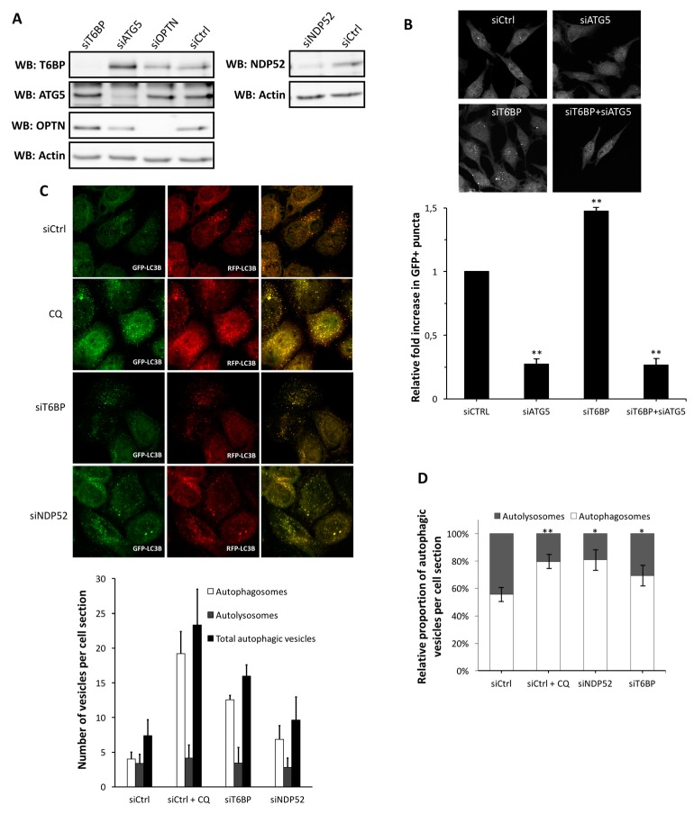
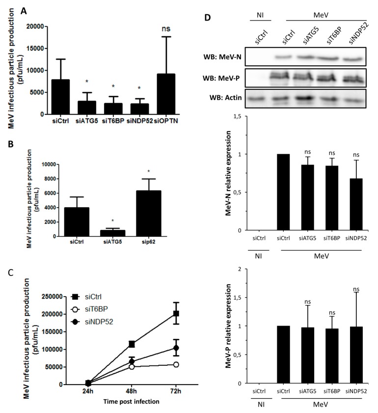
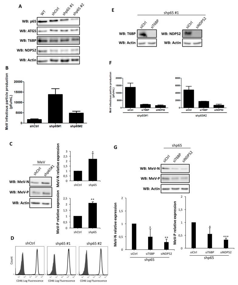
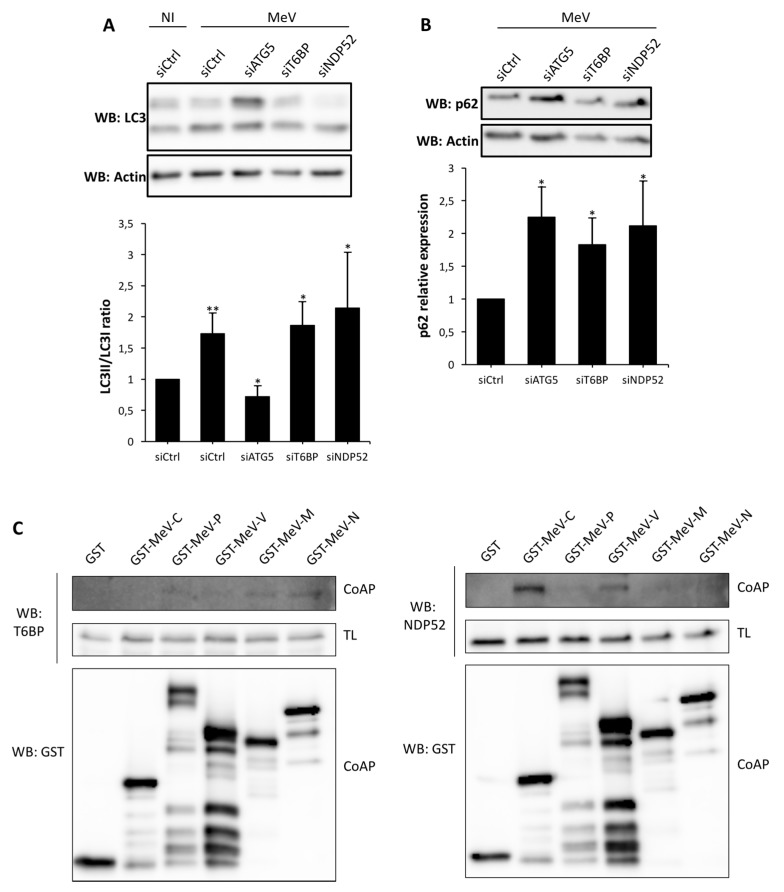
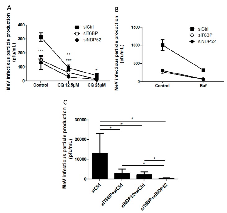
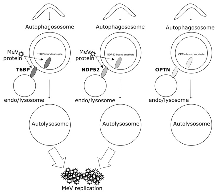
Autophagy is a potent cell autonomous defense mechanism that engages the lysosomal pathway to fight intracellular pathogens. Several autophagy receptors can recognize invading pathogens in order to target them towards autophagy for their degradation after the fusion of pathogen-containing autophagosomes with lysosomes. However, numerous intracellular pathogens can avoid or exploit autophagy, among which is measles virus (MeV). This virus induces a complete autophagy flux, which is required to improve viral replication. We therefore asked how measles virus interferes with autophagy receptors during the course of infection. We report that in addition to NDP52/CALCOCO₂ and OPTINEURIN/OPTN, another autophagy receptor, namely T6BP/TAXIBP1, also regulates the maturation of autophagosomes by promoting their fusion with lysosomes, independently of any infection. Surprisingly, only two of these receptors, NDP52 and T6BP, impacted measles virus replication, although independently, and possibly through physical interaction with MeV proteins. Thus, our results suggest that a restricted set of autophagosomes is selectively exploited by measles virus to replicate in the course of infection.

Keywords: autophagosome; autophagy receptor; maturation; measles virus.


Conflict of interest statement

Authors declare no conflict of interest.

Figures

Figure 1
Figure 1
T6BP function in autophagosome maturation. (A) HeLa cells transfected with the indicated short interfering RNAs (siRNAs) for 48 h, were lysed, and the expression of relevant proteins was probed by Western blotting; (B) GFP-LC3 HeLa cells were transfected or co-transfected with the indicated siRNAs for 48 h and fixed for analysis by confocal microscopy. Representative profiles are shown along with a graph expressing the relative fold induction of the dot number compared with control cells; (C) mRFP-GFP-LC3 HeLa cells were transfected with the indicated siRNAs for 48 h and were treated or not treated during the last 2 h of culture with chloroquine. Representative profiles of autophagosomes (RFP+GFP+ dots) and autolysosomes (RFP+GFP dots) per cell section assessed by confocal microscopy are shown and were quantified. Results are expressed as absolute numbers of individual vesicles (total autophagic vesicles = all RFP+ dots); (D) Results in (C) are shown as the percentage of total autophagic vesicles; (B,C) were each carried out three times in duplicates. GFP: green fluorescent protein; RFP: red fluorescent protein; WB: Western blot; Ctrl: control; CQ: chloroquine.
Figure 2
Figure 2
Involvement of autophagy receptors in measles virus (MeV) replication. (A,B) HeLa cells were transfected with the indicated siRNAs for 48 h, then infected with MeV (multiplicity of infection (MOI) 0.1). 48 h post infection infectious virus particles were titrated by a plaque assay; (C) HeLa cells were transfected with the indicated siRNA for 48 h, then infected with MeV (MOI 1). One, two, or three days post infection, infectious virus particles were titrated by a plaque assay; (D) Cells were treated as in (A). Expression of measles virus N and P proteins was assessed by Western blotting. Representative results are shown and are accompanied by a graph representing the intensity of MeV-N and MeV-P expression over Actin normalized to Control condition.(A,B,D error bars and mean ± SD are from three independent experiments; C is one experiment representative of two independent ones carried out in duplicates). NI: non infected
Figure 3
Figure 3
NF-κB-independent role of T6BP and NDP52 in MeV replication. (A) p65/RelA-expressing HeLa cells and shControl-expressing HeLa cells were transfected with the indicated siRNAs for 48 h, then lysed, and the expression of relevant proteins was probed by Western blot; (B) p65/RelA-expressing HeLa cells and shControl-expressing cells were infected with MeV (MOI 0.1). 48 h post infection, infectious virus particles were titrated by a plaque assay; (C) Cells from (B) were lysed 48 h post infection. Expression of measles virus N and P proteins were assessed by Western blotting. Representative results from shp65#1 are shown and are accompanied by a graph representing the intensity of MeV-N and MeV-P expression over Actin normalized to shControl-expressing cells condition. Means ± SD of four independent experiments are represented (two with the shp65#1 cell line and two with the shp65#2 cell line); (D) p65/RelA-expressing HeLa cells and shControl-expressing HeLa cells were stained for CD46 expression and analyzed by flow cytometry; grey histograms = isotype control, white histograms = CD46 labelling. (EG) p65/RelA-expressing HeLa cells were treated with indicated siRNAs for 48 h; (E) Cells were lysed and the expression of relevant proteins was probed by Western blotting. Results regarding cell line shp65 #1 are represented. Similar results were obtained with shp65 #2. Cells were infected with MeV (MOI 0.1) and 48 h post infection, infectious virus particles were titrated by a plaque assay (F) or lysed; (G) Expression of measles virus MeV-N and MeV-P proteins was assessed by Western blotting. Representative results are shown and are accompanied by a graph representing the intensity of measles proteins expression over Actin normalized to control siRNA condition; (B,F) Means ± SD of one representative experiment out of two independent ones carried out with each shp65/RelA-expressing cell line in duplicates; (G) Means ± SD of four independent experiments.
Figure 4
Figure 4
MeV protein interactions with NDP52 and T6BP. (A) HeLa cells were transfected with the indicated siRNAs for 48 h, and infected or not infected with MeV (MOI 0.1). 48 h post infection, cells were lysed, and anti-LC3 and anti-Actin Western blots were performed. Representative results are shown along with a graph representing the intensity of LC3 II/LC3 I bands normalized to the uninfected control condition; (B) HeLa cells were transfected with the indicated siRNAs for 48 h and infected with MeV (MOI 0.1). 48 h post infection, cells were lysed, and anti-p62 and anti-Actin Western blots were performed. Representative results are shown along with a graph representing the intensity of p62/Actin bands normalized to the control condition; (A,B) Means ± SD of three independent experiments are represented; (C) Cells were transfected with vectors encoding the indicated viral protein. Two days later, cells were lysed and GST-tagged proteins precipitated and proteins were blotted for endogenous T6BP or NDP52 as indicated. Co-AP: co affinity precipitation; TL: total lysate, GST: glutathione S-transferase.
Figure 5
Figure 5
Autophagosome maturation and MeV replication. (A,B) HeLa cells were transfected with the indicated siRNAs for 48 h. Cells were then simultaneously infected with MeV (MOI 0.1) and treated or not treated with 25 µM or 12.5 µM of Chloroquine (A) or 25 nM of Bafilomycin A1 (Baf A1) (B) 48 h post infection and drug treatment, infectious virus particles were titrated by a plaque assay; (C) HeLa cells were transfected or co-transfected with the indicated siRNAs for 48 h. Cells were infected with MeV (MOI 0.1) and 48 h post infection, infectious virus particles were titrated by a plaque assay. Means ± SD of three independent experiments performed in duplicates are represented; (A,C), means ± SD of three to four independent experiments performed in duplicates; (B) means ± SD of one representative experiment out of four independent ones.
Figure 6
Figure 6
Schematic model of the interplay of autophagy receptors with MeV replication. NDP52, OPTN, and T6BP could all play a dual function in autophagy: to target selective substrates towards autophagosomes and to regulate substrate-containing autophagosome maturation (which could be those for which they targeted selective substrates), for an efficient degradation. Only autophagosome maturated via an NDP52 or T6BP pathway are exploited by MeV to improve its replication. Such exploitation could occur via the usage of each receptor for their functions in the targeting and/or the maturation processes (dashed arrows).

References

    1. Boya P., Reggiori F., Codogno P. Emerging regulation and functions of autophagy. Nat. Cell Biol. 2013;15:713–720. doi: 10.1038/ncb2788. - DOI - PMC - PubMed
    1. Gomes L.C., Dikic I. Autophagy in antimicrobial immunity. Mol. Cell. 2014;54:224–233. doi: 10.1016/j.molcel.2014.03.009. - DOI - PubMed
    1. Gregoire I.P., Richetta C., Meyniel-Schicklin L., Borel S., Pradezynski F., Diaz O., Deloire A., Azocar O., Baguet J., Le Breton M., et al. IRGM is a common target of RNA viruses that subvert the autophagy network. PLoS Pathog. 2011;7:e1002422. doi: 10.1371/journal.ppat.1002422. - DOI - PMC - PubMed
    1. Richetta C., Grégoire I.P., Verlhac P., Azocar O., Baguet J., Flacher M., Tangy F., Rabourdin-Combe C., Faure M. Sustained autophagy contributes to measles virus infectivity. PLoS Pathog. 2013;9:e1003599. doi: 10.1371/journal.ppat.1003599. - DOI - PMC - PubMed
    1. Moss W.J., Griffin D.E. Measles. Lancet. 2012;379:153–164. doi: 10.1016/S0140-6736(10)62352-5. - DOI - PubMed

MeSH terms

LinkOut - more resources