Review
. 2017 May 22;9(5):125.
doi: 10.3390/v9050125.

A Role for the Host DNA Damage Response in Hepatitis B Virus cccDNA Formation-and Beyond?

Affiliations
Review

A Role for the Host DNA Damage Response in Hepatitis B Virus cccDNA Formation-and Beyond?

Sabrina Schreiner et al. Viruses. .

Abstract

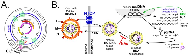
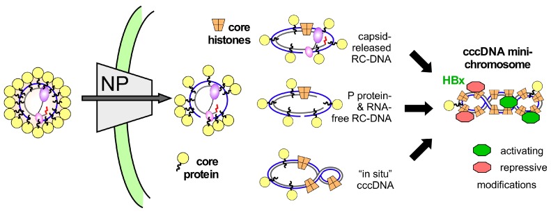
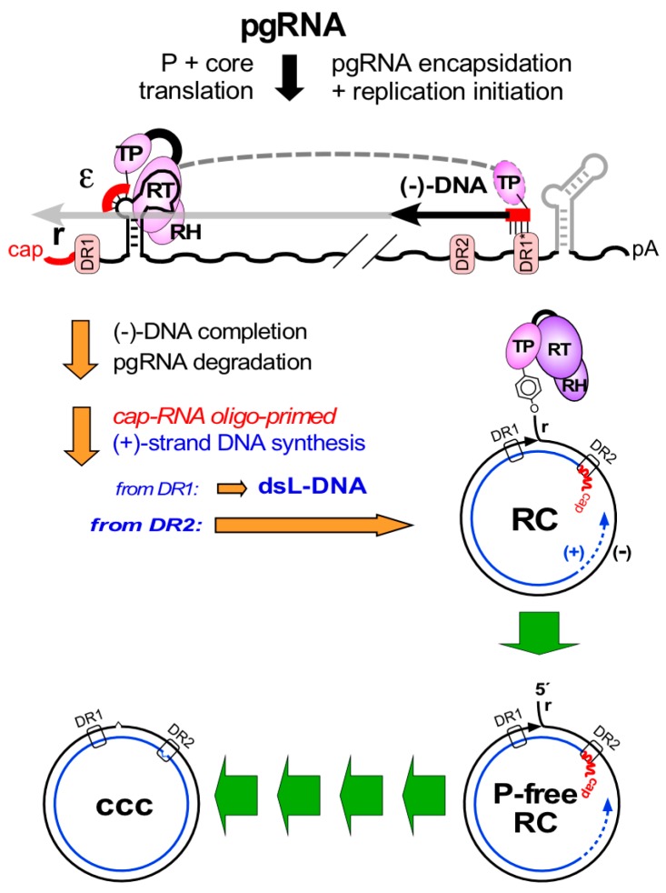
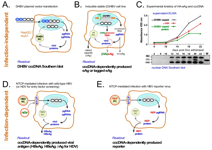
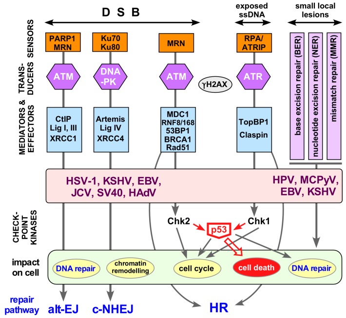
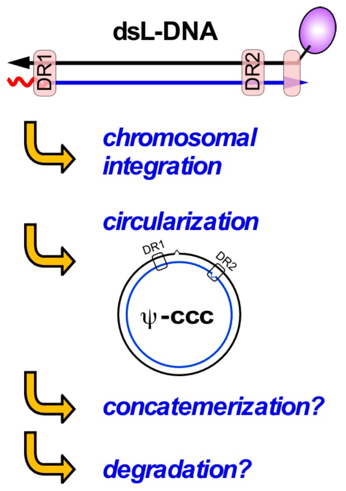
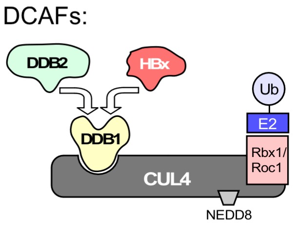
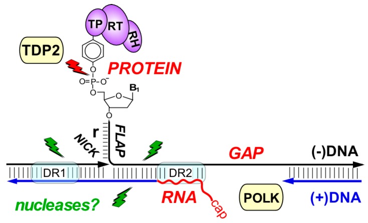
Chronic hepatitis B virus (HBV) infection puts more than 250 million people at a greatly increased risk to develop end-stage liver disease. Like all hepadnaviruses, HBV replicates via protein-primed reverse transcription of a pregenomic (pg) RNA, yielding an unusually structured, viral polymerase-linked relaxed-circular (RC) DNA as genome in infectious particles. Upon infection, RC-DNA is converted into nuclear covalently closed circular (ccc) DNA. Associating with cellular proteins into an episomal minichromosome, cccDNA acts as template for new viral RNAs, ensuring formation of progeny virions. Hence, cccDNA represents the viral persistence reservoir that is not directly targeted by current anti-HBV therapeutics. Eliminating cccDNA will thus be at the heart of a cure for chronic hepatitis B. The low production of HBV cccDNA in most experimental models and the associated problems in reliable cccDNA quantitation have long hampered a deeper understanding of cccDNA molecular biology. Recent advancements including cccDNA-dependent cell culture systems have begun to identify select host DNA repair enzymes that HBV usurps for RC-DNA to cccDNA conversion. While this list is bound to grow, it may represent just one facet of a broader interaction with the cellular DNA damage response (DDR), a network of pathways that sense and repair aberrant DNA structures and in the process profoundly affect the cell cycle, up to inducing cell death if repair fails. Given the divergent interactions between other viruses and the DDR it will be intriguing to see how HBV copes with this multipronged host system.

Keywords: DNA damage response; DNA repair; HBV cure; HBV minichromosome; cccDNA; hepatitis B virus.


Conflict of interest statement

The authors declare no conflict of interest.

Figures

Figure 1
Figure 1
Molecular basics of Hepatitis B Virus (HBV). (A) HBV genome organization. Shown from inside to outside are the open reading frames (ORFs) with their designations; the relaxed circular DNA (RC-DNA) strands with the relative positions of direct repeat 1 (DR1), DR2, enhancer I, enhancer II and the four internal promotors (green arrows); and the transcripts with their staggered 5′ ends (arrowheads) and common 3′ polyA ends. ε denotes the RNA stem-loop on pregenomic RNA (pgRNA) that directs co-encapsidation of pgRNA and P protein and protein-primed replication initiation; (B) Simplified genome replication cycle. Virus entry is mediated by binding of L protein’s PreS1 domain to Na+-taurocholate cotransporting polypeptide (NTCP) [15,16] and additional entry factors (not shown) such as glypican 5 [17]. Nucleocapsids stripped from the envelope transport the P protein-linked RC-DNA to the nucleus where conversion into covalently closed circular DNA (cccDNA) takes place. cccDNA serves as template for the various transcripts, including pgRNA from which core protein and P protein are translated. Via P protein binding to ε, pgRNA is encapsidated (“immature” nucleocapsid) and reverse transcribed into new RC-DNA (“mature” nucleocapsid); this step is inhibited by therapeutic nucleos(t)ide analogs (NAs). Mature progeny nucleocapsids can be enveloped and secreted, or retransport the new RC-DNA to the nucleus to increase cccDNA copy number (“intracellular recycling”). Subgenomic (sg) RNAs act as mRNAs for the envelope proteins and hepatitis B virus X protein HBx which stimulates transcriptional activity of cccDNA (green arrow). Translation of the precore RNA which includes the preC start codon yields precore protein which is processed and secreted as HBeAg.
Figure 2
Figure 2
A speculative model for HBV cccDNA minichromosome formation. Interactions at the nuclear pore (NP) cause disintegration of the nucleocapsid structure [18]; however, due to the core protein’s nucleic acid binding C-terminal domain (CTD) (wiggly lines emanating from the yellow spheres symbolizing core protein) not all core protein subunits may be immediately stripped from the RC-DNA. Loading with histones could thus initiate on complexes with still bound P protein and largely unprocessed RC-DNA, or any time later when P protein is released and one or both DNA strands are freshly ligated (termed “in situ” cccDNA in Figure 2). While eventually most molecules will be covalently closed and fully chromatinized, activating and repressive modifications (symbolized by the green and red objects), modulatable by HBx, may be added before this state is reached. In reality, it is likely that on a single cccDNA minichrosome either activating or repressive marks dominate.
Figure 3
Figure 3
The hepadnaviral genome life-cycle. Protein-primed reverse transcription is initiated by P protein binding to the ε stem-loop on pgRNA, leading to a short ε-templated DNA oligo whose 5′ terminal nt is covalently linked to a Tyr residue in P protein’s terminal protein (TP) domain. Translocation to an acceptor at DR1* allows its extension into slightly overlength minus-strand DNA (carrying the ”r” redundancy), with concomitant pgRNA degradation, except for the capped 5′ terminal end that serves as plus-strand DNA primer. Direct extension from DR1 on yields double stranded linear DNA (dsL-DNA); RC-DNA formation requires primer transfer to DR2 plus an additional template switch (not shown). These steps establish the unusual features of RC-DNA, with non-DNA moieties on both 5′ termini, an overlength minus-strand and an incomplete plus-strand. For cccDNA formation, all peculiarities on RC-DNA must be fixed, both strands must gain exactly unit-length, and the ends must be ligated. The multistep nature of the process is symbolized by the multiple green arrows. One of the predicted intermediates is RC-DNA from which P protein has been released (P-free RC); whether this is the first intermediate as depicted is not known.
Figure 4
Figure 4
A detour to the bench—suitability of different exonucleases for selective degradation of non-cccDNA forms. (A) Model substrates. A 3.2 kb plasmid carrying 800 bp of HBV sequence (lane ccc) was linearized (lane lin) with Bsp QI or nicked (lane RC) with nickase Nt.Bsp QI (both NEB); (B) Differential sensitivity towards Plasmid-Safe DNase (PSD) and T7 and T5 exonuclease (T7exo, T5exo). Plasmid RC-DNA and cccDNA were mixed to contain per 50 µL reaction 2 ng or 200 pg of each plasmid DNA plus 4 µg genomic DNA (gDNA) from Huh7 cells (equivalent to 0.6 × 10⁶ cells). After adjusting buffer conditions as recommended by the nuclease manufacturers reactions were supplemented with 10 U of PSD (Epicentre; 1× Plasmid-Safe reaction buffer with 1 mM ATP), or T7 or T5 exonuclease (both NEB; 1× NEBuffer 4) and incubated at 37 °C (PSD, T5 exo) or 25 °C (T7 exo) for 30 or 120 min. After agarose gel electrophoresis gDNA was detected by ethidium bromide staining (bottom panels), HBV plasmid forms by Southern blotting using a 32P-labeled HBV DNA probe. M, 50 pg each of the ccc, RC and linear form of the HBV plasmid; (C) Detailed time course for T5 exonuclease digestion. An ideal nuclease treatment would completely digest all non-cccDNA forms while fully preserving cccDNA; T5 exonuclease came closest to the first but not to the second criterion.
Figure 5
Figure 5
Surrogate models to overcome low production and poor specific detection of HBV cccDNA. (A) Transient transfection of DHBV expression vectors into human hepatoma cells. DHBV produces much more cccDNA than HBV in the same human hepatoma cells [36]. Transfected plasmid can be selectively digested using the bacterial methylation-dependent restriction enzyme Dpn I while cccDNA amounts suffice for Southern blot detection; (B) Stable, inducibly HBV or DHBV producing hepatoma cell lines. Such cell lines contain a Tet-responsive transactivator (tTA) and an integrated virus expression cassette in which pgRNA is transcribed from an inducible heterologous promoter (e.g., TRE) which does not direct transcription of precore RNA; hence no precore protein or HBeAg is produced. Formation of cccDNA enables precore RNA and precore/HBeAg synthesis. However, specificity of HBeAg detection is limited by crossreactivity with core protein from released naked capsids; this has recently been improved by adding short tags, e.g., HA, specifically to HBeAg; (C) Synchronous kinetics of secreted HA-DHBeAg and cccDNA production in an inducible DHBV HepG2 line encoding HA-tagged DHBeAg. Expression of pgRNA was induced by Dox withdrawal; at the indicated time points intact DHBV capsids, DHBV capsid protein (as present in DHBeAg and disassembled capsids) and HA-tag in the culture supernatants were monitored by ELISA; in parallel, nuclear DNAs were analyzed by Southern blotting; (D) Infection-dependent cccDNA formation with wild-type HBV. Productive infection of NTCP-expressing cells depends on prior cccDNA formation, resulting in generation of viral antigens; hence HBsAg and HBeAg can serve as surrogate markers for cccDNA production. Interference with other infection steps would cause the same readout; entry-specific factors may be identified by using HDV [17] which shares only the early infection steps with HBV; (E) Improved detection of infection via HBV reporter vectors. Easily detectable reporters (REP) encoded by modified HBVs and expressed in a cccDNA-dependent fashion would allow more sensitive and better quantifiable monitoring. As yet, however, such HBV vectors are much less advanced than for other virus families.
Figure 6
Figure 6
The host DNA damage repair response and viral interference. Double-strand DNA breaks (DSBs), exposed single stranded DNA (ssDNA) at collapsed replication forks and various kinds of local DNA lesions are detected by sensors that mark the site of damage. Transducers, and mediator plus effector proteins transmit and amplify the signal throughout the cell, resulting in a huge influx of factors to repair damage and remodel chromatin. The key apical transducer kinases are ATM (Ataxia telangiectasia mutated), ATR (Ataxia telangiectasia and Rad3-related), and DNA-PK (DNA-dependent protein kinase). These Ser/Thr kinases regulate DNA replication, DNA repair, cell-cycle checkpoint control (e.g., via Chk1, Chk2), and if necessary cell death (e.g., via p53) by recruitment of specific effector proteins. A hallmark of the DNA damage respone (DDR) is phosphorylation of histone H2AX to generate γH2AX, and the formation of large γH2AX foci around the site of damage. c-NHEJ and alt-EJ are error-prone and always active; high-fidelity repair via homologous recombination (HR) requires an intact sister chromatide as template and thus is largely restricted to the S and G2 phases of the cell cycle. More and more viruses are known to exhibit a complex relationship with the DDR [101,106] to maintain efficient viral replication and onset of gene expression. Only some well established DNA virus examples are shown (HSV-1, herpes simplex virus type 1; EBV, Epstein-Barr-virus; KSHV; Kaposi’s sarcoma herpesvirus; JCV, JC virus; SV40, simian virus 40; HPV, human papillomavirus; MCPyV; Merkel cell polyomavirus; HAdV, human adenovirus) but RNA viruses are joining the list [99]. See text for further details. Abbreviations: 53BP1, P53-binding protein 1; BRCA1, Breast cancer susceptibility gene 1; Chk, Checkpoint kinase; CtIP, C-terminal interacting protein; DSB, double-strand DNA break; MDC1, Mediator of DNA damage checkpoint; PARP1, poly ADP ribose polymerase 1; RNF, Ring finger protein; TopBP1, topoisomerase-IIbeta-binding protein 1; XRCC, X-ray repair complementing defective repair in Chinese hamster cells 1.
Figure 7
Figure 7
Double-strand linear (dsL) DNA—a free-ended double-strand DNA break mimic? Hepadnaviral dsL-DNA is a common replication by-product from in-situ priming of plus-strand DNA. Like other linear DNAs dsL-DNA likely evokes repair responses that remove the free ends, e.g., by chromosomal integration, circularization (as for retroviral 2LTR-circles), concatemerization and/or exonucleolytic degradation. Circularization appears to occur mainly via error-prone non-homologous end-joining, yielding often defective cccDNA-like molecules (Ψ-cccDNA). Concatemerization and degradation via exonucleases have not explicitly been demonstrated.
Figure 8
Figure 8
HBx-DDB1 interaction does not necessarily indicate a direct connection to DNA repair. UV-damaged DNA binding protein 1 (DDB1) was reported early on as an HBx interactor. However, different from what the name implies DDB1 does not directly bind to damaged DNA; this function is taken by the distinct DDB2 protein. DDB1’s major function is that of an adaptor in the E3 ubiquitin ligase CRL4, comprising the cullin 4 (CUL4) scaffold protein, a RING finger domain protein (Rbx1 or Roc1) which mediates binding of a ubiquitin conjugating E2 enzyme, and a regulatory site for modification with NEDD8. For ubiquitylation of specific target proteins, DDB1 must interact with DDB1-CUL4 associated factors (DCAFs) which act as substrate receptors. DDB2 is one of multiple cellular DCAFs while HBx is a viral DCAF [31,32,33]. As HBx binds to the same site on DDB1 as cellular DCAFs do [162] their binding is mutually exclusive.
Figure 9
Figure 9
Summary of hepadnaviral RC-DNA as a multi-target DNA repair substrate. Close-up of the molecular peculiarities in RC-DNA. The unusual features of RC-DNA concentrate on a small region encompassing direct repeats DR1 and DR2. P protein is linked to the 5′ end of minus-strand DNA through a tyrosyl-DNA phosphodiester bond; the small “r” redundancy forms a flap preceded by a nick. The incomplete plus-strand starts with RNA and leaves a gap that makes the opposite minus-strand single-stranded. The host enzyme TDP2 can cleave the tyrosyl-DNA-phosphodiester bond to release P protein [49] yet as in repair of cellular protein-DNA adducts alternative, probably nucleolytic repair pathways are likely to exist (green lightning symbols). Translesion DNA polymerase κ (POLK) is thought to fill-in the gap in plus-strand DNA [46] but can probably also be substituted for by other repair polymerases. Factors required for the other RC-DNA modifications have not yet been identified.

References

    1. Trepo C., Chan H.L., Lok A. Hepatitis B virus infection. Lancet. 2014;384:2053–2063. doi: 10.1016/S0140-6736(14)60220-8. - DOI - PubMed
    1. Stanaway J.D., Flaxman A.D., Naghavi M., Fitzmaurice C., Vos T., Abubakar I., Abu-Raddad L.J., Assadi R., Bhala N., Cowie B., et al. The global burden of viral hepatitis from 1990 to 2013: Findings from the Global Burden of Disease Study 2013. Lancet. 2016;388:1081–1088. doi: 10.1016/S0140-6736(16)30579-7. - DOI - PMC - PubMed
    1. WHO Fact sheet 297 Cancer 2017. Feb, 2017.
    1. Götte M., Feld J.J. Direct-acting antiviral agents for hepatitis C: Structural and mechanistic insights. Nat. Rev. Gastroenterol. Hepatol. 2016;13:338–351. doi: 10.1038/nrgastro.2016.60. - DOI - PubMed
    1. Moradpour D., Grakoui A., Manns M.P. Future landscape of hepatitis C research—Basic, translational and clinical perspectives. J. Hepatol. 2016;65:S143–S155. doi: 10.1016/j.jhep.2016.07.026. - DOI - PubMed

Publication types

MeSH terms