Review
. 2017 May 22:46:175-198.
doi: 10.1146/annurev-biophys-051013-022942.

What Do Structures Tell Us About Chemokine Receptor Function and Antagonism?

Affiliations
Review

What Do Structures Tell Us About Chemokine Receptor Function and Antagonism?

Irina Kufareva et al. Annu Rev Biophys. .

Abstract

Chemokines and their cell surface G protein-coupled receptors are critical for cell migration, not only in many fundamental biological processes but also in inflammatory diseases and cancer. Recent X-ray structures of two chemokines complexed with full-length receptors provided unprecedented insight into the atomic details of chemokine recognition and receptor activation, and computational modeling informed by new experiments leverages these insights to gain understanding of many more receptor:chemokine pairs. In parallel, chemokine receptor structures with small molecules reveal the complicated and diverse structural foundations of small molecule antagonism and allostery, highlight the inherent physicochemical challenges of receptor:chemokine interfaces, and suggest novel epitopes that can be exploited to overcome these challenges. The structures and models promote unique understanding of chemokine receptor biology, including the interpretation of two decades of experimental studies, and will undoubtedly assist future drug discovery endeavors.

Keywords: G protein–coupled receptor; allostery; crystallography; druggability; molecular modeling; receptor activation.


Figures

Figure 1
Figure 1. Topology and oligomerization behavior of chemokines
(a–b) Typical structures of a CC (a) and CXC (b) chemokines. (c–d) Typical dimerization geometry of CC (c) and CXC (d) chemokines. (e) CC chemokines bind GAGs through extensive positively charged interfaces formed through chemokine oligomerization. The oligomers are formed by polymerization of dimers that have a geometry consistent with (c).
Figure 2
Figure 2. Chemokine interaction with receptors and other binding partners
(a) Canonical two-site model of receptor:chemokine recognition. (b-c) Structures of receptor:chemokines complexes solved in 2015 (CXCR4:vMIP-II and US28:CX3CL1) are consistent with the two-site model. Important basic residues on the chemokines are highlighted in blue. The chemokine N-terminus is highlighted in green. The Pro of the receptor Pro-Cys motif is colored cyan and packs up against the conserved disulfide of the chemokine (yellow surface). Proximal sulfotyrosine residues from the receptor N-termini were modeled and are shown with the sulfate colored red and yellow. (d-e) The CXC chemokine dimerization geometry (d) is closely mimicked by the hypothetical interaction of distal receptor N-terminus with the β1-strand of the chemokine in the modeled ACKR3:CXCL12 complex (e). The prediction is supported by the radiolytic footprinting data. (f) The binding interface of CXCL1 consisting of the proximal N-terminus of the chemokine and its N-loop/40s loop groove is shared by GAGs and the receptor. (g-j) CC chemokine dimers and pathogen chemokine-binding proteins share receptor binding interfaces and geometry.
Figure 3
Figure 3. Insights into chemokine receptor activation
(a-f) Residues critical for CXCL12-induced activation of CXCR4 were identified and mapped onto an inactive and active state model of CXCR4:CXCL12 complex. (a-b) and (d-f) Five structural and functional layers are shown in the context of the full structure (center) and enlargements: (a) chemokine engagement (blue), (b) signal initiation (green), (d) signal propagation (yellow), (e) microswitch activation (red) and (f) G protein coupling (purple). Predicted residue conformations in the inactive and active state are shown in lighter and darker colors, respectively. (c) The proposed geometry of the interaction between the CXCL12 distal N-terminus (a critical signaling domain, shown in black) and chemokine engagement (blue)/signal initiation (green) residues of CXCR4. (g) Radiolytic footprinting mapping of residue solvent exposure in the atypical chemokine receptor ACKR3. Residues protected by the chemokine (as compared to a small-molecule bound state) are shown in shades of red: these residues are mostly located in the N-terminus and the extracellular loops of the receptor. On the contrary, numerous residues within the transmembrane domain become less protected in the chemokine-bound state; these residues report on increased solvent exposure of the TM core due to conformational changes upon chemokine-induced full activation of ACKR3.
Figure 4
Figure 4. The allosteric nature of chemokine receptor activation and inhibition
(a) Extracellular chemokine binding translates into intracellular G protein coupling, and communication also works in reverse with G protein binding enhancing the affinity of chemokines. (b) Orthosteric antagonists directly block binding of chemokines in the CRS2 ligand binding pocket. (c-e) Apparently allosteric behavior of chemokine receptor antagonists can have multiple explanations including binding at a site distinct from the chemokine (c), occupying a part of the extensive chemokine interaction interface while allowing for a ternary complex formation (d), or binding to a distinct sub-population of receptors (e). The latter can happen in heterogeneous receptor populations where the G protein-coupled sub-population preferentially binds chemokine agonist while the uncoupled sub-population binds small molecule antagonists.
Figure 5
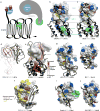
Figure 5. Structural basis of small molecule antagonism of chemokine receptors
(a) Receptors (grey) interact with chemokines (magenta) via extensive interfaces with numerous polar contacts (cyan); the interaction is additionally reinforced with the flexible N-terminus of the receptor (CRS1) essentially wrapping around the chemokine. Like most protein:protein interactions, chemokine:receptor interactions are conceptually difficult to inhibit with small molecules. (b-d) Structures of chemokine receptors with small molecule antagonists: (b) CCR5:maraviroc (c) CXCR4:IT1t, and (d) CCR2 in a ternary complex with BMS-681 and CCR2-RA-[R]. Each molecule explores a unique non-polar subpocket (highlighted in yellow) within the overall large and polar (i.e. poorly druggable) binding pocket. (e) All three crystallized orthosteric small molecule antagonists of chemokine receptors demonstrate low degree of enclosure and high degree of solvent exposure. (f) This is in stark contrast with small molecule antagonists of non-chemokine GPCRs. (g) The crystallized allosteric antagonist of CCR2 binds in an intracellular pocket with favorable druggability properties and demonstrates a high degree of enclosure.

References

    1. Alexander-Brett JM, Fremont DH. Dual GPCR and GAG mimicry by the M3 chemokine decoy receptor. The Journal of Experimental Medicine. 2007;204:3157–72. - PMC - PubMed
    1. Andrews G, Jones C, Wreggett KA. An Intracellular Allosteric Site for a Specific Class of Antagonists of the CC Chemokine G Protein-Coupled Receptors CCR4 and CCR5. Molecular Pharmacology. 2008;73:855–67. - PubMed
    1. Baggiolini M. Chemokines and leukocyte traffic. Nature. 1998;392:565–68. - PubMed
    1. Ballesteros JA, Weinstein H. Integrated methods for the construction of three-dimensional models and computational probing of structure-function relations in G protein-coupled receptors. In: Sealfon SC, editor. Methods in Neurosciences: Receptor Molecular Biology. San Diego: Academic Press; 1995. pp. 366–428.
    1. Bertini R, Allegretti M, Bizzarri C, Moriconi A, Locati M, et al. Noncompetitive allosteric inhibitors of the inflammatory chemokine receptors CXCR1 and CXCR2: Prevention of reperfusion injury. Proceedings of the National Academy of Sciences of the United States of America. 2004;101:11791–96. - PMC - PubMed