Review
. 2016 Oct 14:2:17-35.
doi: 10.1146/annurev-vision-111815-114638.

Experience-Dependent Structural Plasticity in the Visual System

Affiliations
Review

Experience-Dependent Structural Plasticity in the Visual System

Kalen P Berry et al. Annu Rev Vis Sci. .

Abstract

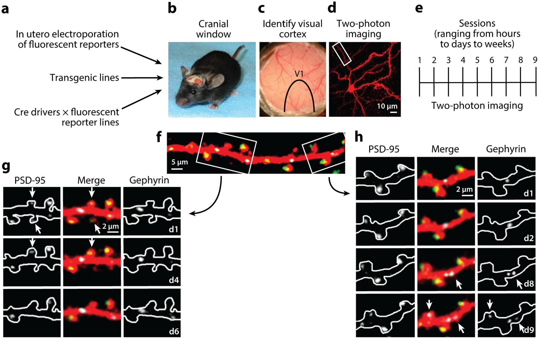
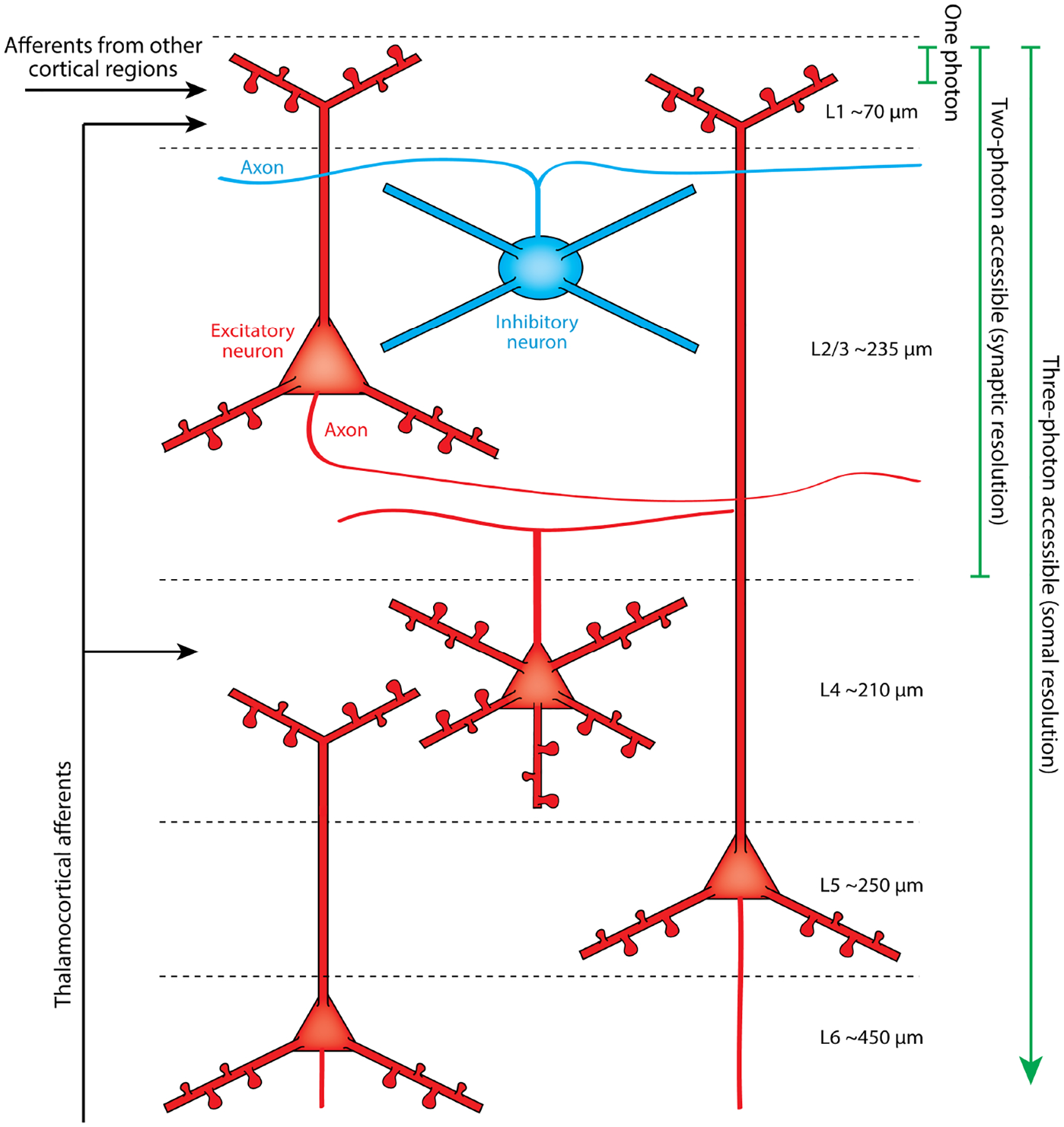
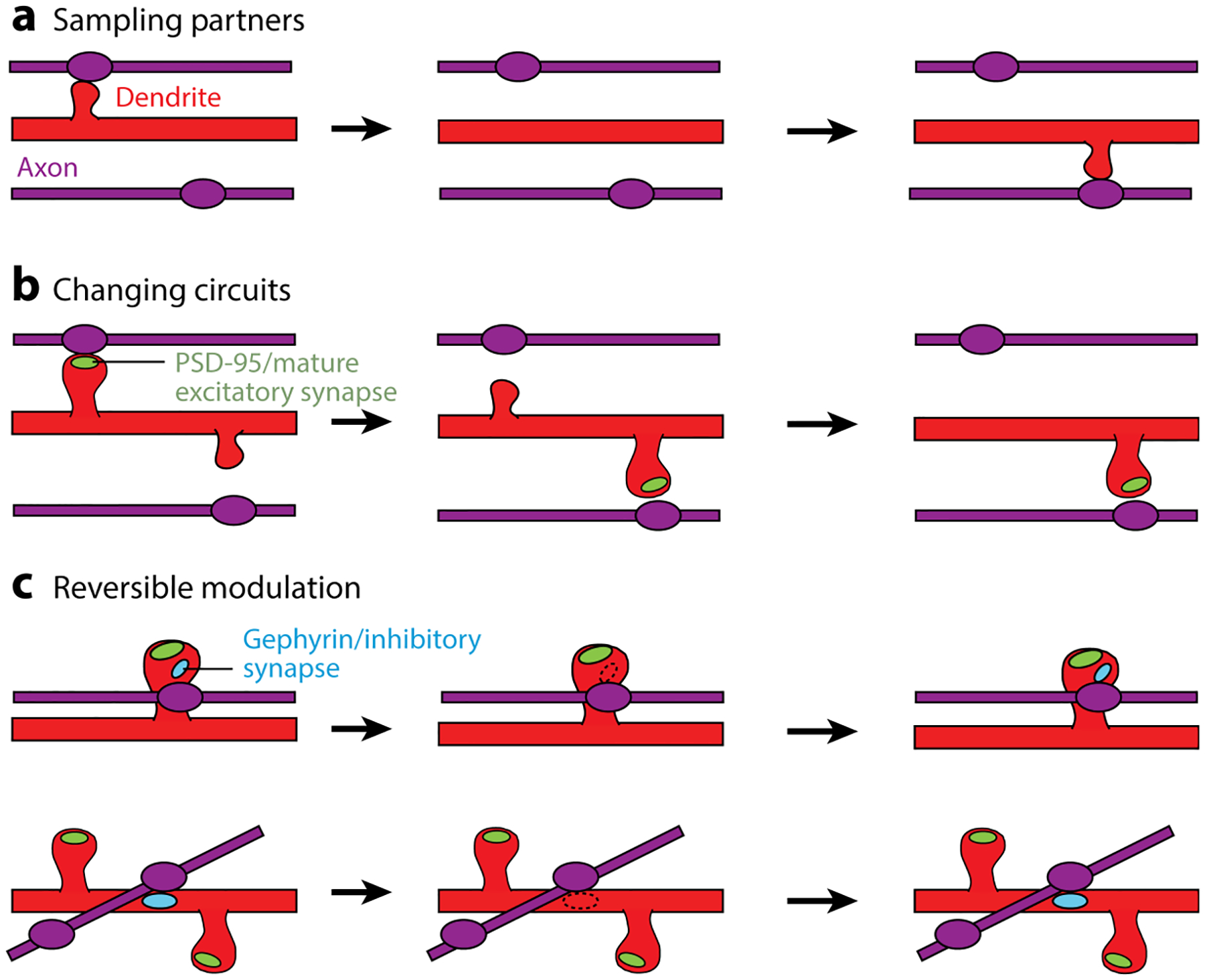
During development, the environment exerts a profound influence on the wiring of brain circuits. Due to the limited resolution of studies in fixed tissue, this experience-dependent structural plasticity was once thought to be restricted to a specific developmental time window. The recent introduction of two-photon microscopy for in vivo imaging has opened the door to repeated monitoring of individual neurons and the study of structural plasticity mechanisms at a very fine scale. In this review, we focus on recent work showing that synaptic structural rearrangements are a key mechanism mediating neural circuit adaptation and behavioral plasticity in the adult brain. We examine this work in the context of classic studies in the visual systems of model organisms, which have laid much of the groundwork for our understanding of activity-dependent synaptic remodeling and its role in brain plasticity.

Keywords: circuit remodeling; retinogeniculate afferents; retinotectal system; structural plasticity; synapse dynamics; visual cortex.


Figures

Figure 1
Figure 1
In vivo two-photon imaging experimental pipeline. (a) Multiple methods for sparsely labeling individual neurons in vivo. (b) Cranial windows are implanted at various times after the mouse is born. (c) Intrinsic signal imaging is used to locate the visual cortex within the cranial window. Blood vessels are used as landmarks to relocate the same cell for multiple imaging sessions. (d) Maximum Z-projection of a cell in the visual cortex. (e) The same cell can be imaged over a range of time periods ranging from hours to days to weeks.(f) Zoomed-in view of boxed region in panel d. Cell fill pseudocolored red, with labeled inhibitory synapses (white), and excitatory synapses (pseudocolored green). (g,h) Examples of dynamic synapses from boxed regions in panel f on the indicated days. Left, middle, and right subpanels show a postsynaptic density protein 95 (PSD-95)-mCherry alone, three-channel merge, and Teal-gephyrin alone, respectively. Arrows denote dynamic synapses. Inhibitory synapses in panel h appear on days 8 and 9 (d8 and d9). The excitatory synapse in panel g disappears on day 4 (d4), and its spine is removed on day 6 (d6).
Figure 2
Figure 2
Cortical circuits accessible through a cranial window. This schematic shows the neuron types accessible in vivo with one-photon, two-photon, or three-photon microscopy. Axons are illustrated as thin lines. One-photon techniques, such as confocal microscopy, cannot image more than a few tens of microns into scattering tissue. Two-photon microscopy can provide high synaptic resolution images throughout 300–400 μm of tissue. Three-photon microscopy can image as deep as a millimeter, and the resolution currently allows cellular but not synaptic imaging. Cortical layer thicknesses according to DeFelipe et al. (2002).
Figure 3
Figure 3
Different logic for excitatory versus inhibitory synaptic changes. (a–c) Schematics illustrating the most prevalent categories of dynamic events for spines without postsynaptic density protein 95 (PSD-95), spines with PSD-95, and inhibitory synapses on dually innervated spine (DiS) and on the shaft. (a) The dynamics of spines without PSD-95 are rapid and sample different locations, potentially testing for different partners.(b) Spines that lose an excitatory synapse are destabilized, whereas those that gain one are stabilized and persist. In both cases, they represent a local rewiring of excitatory circuits by exchanging partners.(c) Inhibitory synapses on the shaft or on DiS are removed and reassembled at stable locations, providing a mechanism for reversible inhibitory modulation of excitatory circuits. Figure adapted from Villa et al. (2016).

References

    1. Ackman JB, Burbridge TJ, Crair MC. 2012. Retinal waves coordinate patterned activity throughout the developing visual system. Nature 490:219–25 - PMC - PubMed
    1. Antonini A, Fagiolini M, Stryker MP. 1999. Anatomical correlates of functional plasticity in mouse visual cortex. J. Neurosci 19:4388–406 - PMC - PubMed
    1. Antonini A, Gillespie DC, Crair MC, Stryker MP. 1998. Morphology of single geniculocortical afferents and functional recovery after reverse monocular deprivation in kitten. J. Neurosci 18:9896–909 - PMC - PubMed
    1. Antonini A, Stryker MP. 1993a. Development of individual geniculocortical arbors in cat striate cortex and effects of binocular impulse blockade. J. Neurosci 13:3549–73 - PMC - PubMed
    1. Antonini A, Stryker MP. 1993b. Rapid remodeling of axonal arbors in the visual cortex. Science 260:1819–21 - PubMed

Publication types

LinkOut - more resources