. 2015 Nov 24:1:263-289.
doi: 10.1146/annurev-vision-082114-035334.

Functional Circuitry of the Retina

Affiliations

Functional Circuitry of the Retina

Jonathan B Demb et al. Annu Rev Vis Sci. .

Abstract

The mammalian retina is an important model system for studying neural circuitry: Its role in sensation is clear, its cell types are relatively well defined, and its responses to natural stimuli-light patterns-can be studied in vitro. To solve the retina, we need to understand how the circuits presynaptic to its output neurons, ganglion cells, divide the visual scene into parallel representations to be assembled and interpreted by the brain. This requires identifying the component interneurons and understanding how their intrinsic properties and synapses generate circuit behaviors. Because the cellular composition and fundamental properties of the retina are shared across species, basic mechanisms studied in the genetically modifiable mouse retina apply to primate vision. We propose that the apparent complexity of retinal computation derives from a straightforward mechanism-a dynamic balance of synaptic excitation and inhibition regulated by use-dependent synaptic depression-applied differentially to the parallel pathways that feed ganglion cells.

Keywords: adaptation; amacrine cell; bipolar cell; ganglion cell; parallel pathways; synaptic depression.


Figures

Figure 1
Figure 1
Retinal circuits. (a) The cellular and synaptic (i.e., plexiform) layers of the retina. Some of the various cell types composing the five classes of neurons are shown: rod and cone photoreceptors, horizontal cells (HCs), ON and OFF cone bipolar cells (BCs), rod BCs, AII and wide-field (WF) amacrine cells (ACs), and ON and OFF ganglion cells (GCs). The ON and OFF BC axon terminals and GC dendrites stratify in separate halves of the inner plexiform layer. (b) Several cell types from panel a, redrawn to illustrate how rod signals pass through the inner retina. Excitatory (+) and inhibitory (−) synapses are shown. A gap junction (denoted by the resistor symbol) allows bidirectional current flow between AII ACs and ON cone BCs. The AII AC splits the ON rod BC signal into ON and OFF components using either electrical (gap junction, ON) or chemical (glycinergic, OFF) synapses. Note that in daylight conditions, cone-mediated drive to the AII influences the OFF pathway as follows: cone → ON cone BC→ AII AC→ OFF BC and GC.
Figure 2
Figure 2
Regional variation in cell density across the retina. (a) Map depicting the gradient of cone opsin coexpression across the mouse retina. In the dorsal/temporal retina, the coexpressing cones, which represent ~90–95% of the cone population, express primarily M opsin, which is most sensitive to green light. In the ventral/nasal retina, the co-expressing cones express primarily S opsin, which is most sensitive to UV light. In between these two extremes, there is a gradient of relative M/S opsin expression. Throughout the retina, approximately 5–10% of cones are genuine S cones and express exclusively S opsin. (b) Map depicting the regional differences in the size and density of two ganglion cell types. In the ventral retina, the W3 ganglion cell type is relatively small, and its mosaic is dense. In the dorsal/temporal retina, the ON αganglion cell type is relatively small, and its mosaic is dense. The contour lines indicate the relative density of either W3 ( green) or ON α(red ) ganglion cell receptive field centers, which is comparable to the dendritic field. The insets (right) show the changes in receptive field size and density for each cell type, illustrating five examples of each cell type within each region.
Figure 3
Figure 3
Synaptic motifs. (a) From the perspective of a bipolar cell (pipette attached), inhibition arising from amacrine cells (ACs) occurs via multiple synaptic motifs. Excitatory (+) and inhibitory (−) synapses are indicated; feedback and feedforward synapses can occur in both ON and OFF systems, and crossover inhibition acts between ON and OFF systems. The illustrated circuit is an ON → OFF inhibitory one, but the opposite pattern (OFF → ON) could also occur. (b) From the perspective of a ganglion cell (GC) (pipette attached), inhibition from ACs occurs via multiple synaptic motifs. This panel follows the same conventions as used in panel a.
Figure 4
Figure 4
Nonlinear subunit receptive field. (a) In one model, bipolar cells have center–surround spatial receptive fields (top), and their synapses are linear (i.e., equal but opposite changes in release rate occur in response to increments or decrements in light intensity). The subunits are integrated by a ganglion cell (GC). The input–output function illustrated in the circle shows the linear relationship between the presynaptic bipolar cell voltage and glutamate release onto the ganglion cell. (b) In a second model, bipolar cell vesicle release is nonlinear because it is rectified (i.e., release can increase but cannot decrease below a low baseline rate). The input–output function illustrated in the circle shows the nonlinear relationship between presynaptic voltage and glutamate release. The circuit is expanded to have an amacrine cell (AC)-mediated surround inhibition that is driven by bipolar cell subunits. Release from the AC is also rectified; thus, inhibition to the bipolar cell terminals and to the GC is rectified. (c) Alternating frames of a grating stimulus viewed by each of two bipolar cell subunits. Each subunit is excited by the bright bar (subunit 1 for frame a, subunit 2 for frame b). (d ) For the linear model in panel a, responses from the bipolar subunits are canceled when summed at the level of the ganglion cell. (e) For the nonlinear model in panel b, stimulation of only the center of a receptive field generates a frequency-doubled response in the ganglion cell when the responses of rectified, nonlinear subunits are summed. When stimulation is extended to the surround, however, AC inhibition cancels the center excitation.
Figure 5
Figure 5
Synaptic mechanism for adaptation. A test pulse is presented in darkness (pulse 1) or added to a background (pulse 2, top trace). The first test pulse evokes a burst of vesicle release. When the background is added, tonic vesicle release increases, and the response to a second test pulse is reduced relative to the first. The readily releasable pool (RRP) of synaptic vesicles is decreased by both the test pulses and the background stimulus. Recovery from depletion of the RRP is relatively slow compared with the decay of release following the test pulse. The result is that a background light depletes the RRP and causes adaptation.
Figure 6
Figure 6
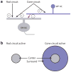
Example of circuit switching. (a) The excitatory input to an ON ganglion cell (GC) is driven by both rod and cone circuits. The rod circuits actually signal via the cone bipolar cell terminal (see Figure 1). The inhibition from the surround is mediated by a wide-field amacrine cell (WF AC) driven exclusively by cone circuits. (b) When the rod circuit is active, the ON GC has a receptive field with an excitatory center component only. When the cone circuit is active, the inhibitory surround component switches on.

References

    1. Applebury ML, Antoch MP, Baxter LC, Chun LL, Falk JD, et al. The murine cone photoreceptor: a single cone type expresses both S and M opsins with retinal spatial patterning. Neuron. 2000;27:513–23. - PubMed
    1. Asari H, Meister M. Divergence of visual channels in the inner retina. Nat Neurosci. 2012;15:1581–89. - PMC - PubMed
    1. Baccus SA, Meister M. Fast and slow contrast adaptation in retinal circuitry. Neuron. 2002;36:909–19. - PubMed
    1. Baccus SA, Olveczky BP, Manu M, Meister M. A retinal circuit that computes object motion. J Neurosci. 2008;28:6807–17. - PMC - PubMed
    1. Baden T, Esposti F, Nikolaev A, Lagnado L. Spikes in retinal bipolar cells phase-lock to visual stimuli with millisecond precision. Curr Biol. 2011;21:1859–69. - PMC - PubMed

LinkOut - more resources