. 2017 May 22;5(1):39.
doi: 10.1186/s40478-017-0443-7.

Multidimensional scaling of diffuse gliomas: application to the 2016 World Health Organization classification system with prognostically relevant molecular subtype discovery

Affiliations

Multidimensional scaling of diffuse gliomas: application to the 2016 World Health Organization classification system with prognostically relevant molecular subtype discovery

Patrick J Cimino et al. Acta Neuropathol Commun. .

Abstract

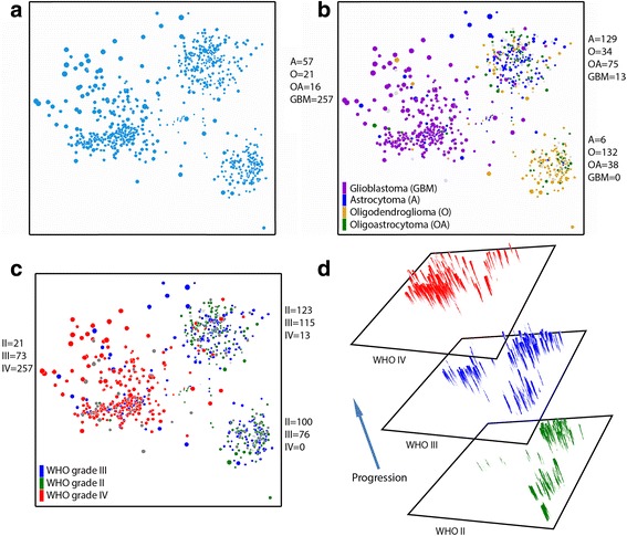
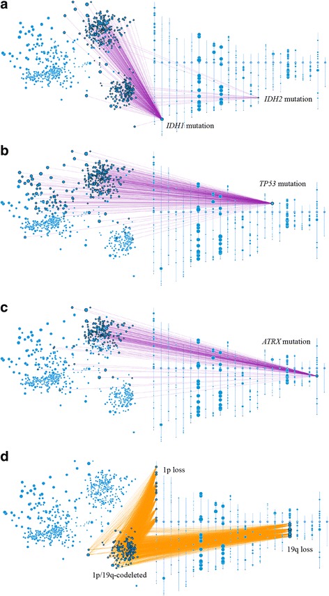
Recent updating of the World Health Organization (WHO) classification of central nervous system (CNS) tumors in 2016 demonstrates the first organized effort to restructure brain tumor classification by incorporating histomorphologic features with recurrent molecular alterations. Revised CNS tumor diagnostic criteria also attempt to reduce interobserver variability of histological interpretation and provide more accurate stratification related to clinical outcome. As an example, diffuse gliomas (WHO grades II-IV) are now molecularly stratified based upon isocitrate dehydrogenase 1 or 2 (IDH) mutational status, with gliomas of WHO grades II and III being substratified according to 1p/19q codeletion status. For now, grading of diffuse gliomas is still dependent upon histological parameters. Independent of WHO classification criteria, multidimensional scaling analysis of molecular signatures for diffuse gliomas from The Cancer Genome Atlas (TCGA) has identified distinct molecular subgroups, and allows for their visualization in 2-dimensional (2D) space. Using the web-based platform Oncoscape as a tool, we applied multidimensional scaling-derived molecular groups to the 2D visualization of the 2016 WHO classification of diffuse gliomas. Here we show that molecular multidimensional scaling of TCGA data provides 2D clustering that represents the 2016 WHO classification of diffuse gliomas. Additionally, we used this platform to successfully identify and define novel copy-number alteration-based molecular subtypes, which are independent of WHO grading, as well as predictive of clinical outcome. The prognostic utility of these molecular subtypes was further validated using an independent data set of the German Glioma Network prospective glioblastoma patient cohort.

Keywords: Astrocytoma; Glioblastoma; Glioma; Isocitrate Dehydrogenase (IDH); Oligodendroglioma; Oncoscape; World Health Organization (WHO).


Figures

Fig. 1
Fig. 1
2D multidimensional scaling plots of TCGA diffuse glioma patients based on genomic data. a Multidimensional scaling shows that there are three main clusters. b 2007 WHO histopathological classification across the three main clusters (number of cases for each cluster is listed). c WHO grades are shown across clusters (number of cases for each cluster is listed). d 3D representation of WHO grading, reflecting progression of each cluster
Fig. 2
Fig. 2
2D diffuse glioma plots with accompanying chromosomal ideograms. a The two main clusters on the right contain mutations in the IDH1 and IDH2 genes, as shown by the purple edges connecting the gene with corresponding patients. The IDH-mutant upper right cluster also carries the majority of (b) TP53 and (c) ATRX gene mutations. d The IDH-mutant lower right cluster contains gliomas that harbor the oligodendroglioma-specific 1p/19q codeletion, as demonstrated by orange edges connecting low level copy loss chromosomal regions with corresponding affected patients. This cluster also contains a majority of the IDH2 mutations
Fig. 3
Fig. 3
2D visualization of the revised 2016 WHO classification of diffuse gliomas. Multidimensional scaling demonstrates three major clusters of diffuse gliomas. The 2007 WHO histopathologic classifiers are heterogeneous and non-specific with regards to the three main clusters. The 2016 WHO classification aligns well with the three major clusters and can be divided into: 1) oligodendroglial tumors, IDH-mutant and 1p/19q-codeleted (WHO grades II–III); 2) astrocytic gliomas/glioblastomas, IDH-mutant (WHO grades II–IV); and 3) astrocytic gliomas/glioblastomas, IDH-wildtype (WHO grades II–IV)
Fig. 4
Fig. 4
Clinical characteristics of TCGA diffuse glioma clusters. a Survival curves from 2007 WHO histopathological classification criteria (A = diffuse astrocytoma, O = oligodendroglioma, OA = oligoastrocytoma, AA = anaplastic astrocytoma, AO = anaplastic oligodendroglioma, AOA = anaplastic oligoastrocytoma, GBM = glioblastoma). b Survival comparison of three main 2D molecular clusters. c-e Age at diagnosis distribution for each cluster. Patients with astrocytic glioma/glioblastoma are older at presentation in the (c) IDH-wildtype cluster than in the (d) IDH mutant cluster. e An apparent bimodal adult age distribution is seen in the oligodendroglioma cluster, with median age of 45 years. f Survival of patients with tumors of the oligodendroglioma cluster stratified by age (<45 versus ≥45 years) is significantly different (p = 0.0033), and comparable to survival stratified by WHO grade (g, h). P values determined using Cox proportional hazard regression
Fig. 5
Fig. 5
Genomic copy number alteration frequency among molecular clusters. Oligodendrogliomas are defined by 1p/19q codeletion and the second most frequent alteration is loss of chromosome 4. The IDH-mutant astrocytic glioma/glioblastoma cluster has several low level copy number alterations, including known astrocytoma-associated alterations such as 9p loss and 19q loss. IDH-wildtype diffuse gliomas have frequent polysomy chromosome 7, chromosome 10 loss, and 9p loss. The IDH-wildtype cluster can be further divided into 3 subgroups (ac). Subgroup A is separated from B and C by either the presence of polysomy chromosome 1 or TP53 mutations. Subgroups B and C are further separated by the presence or absence of polysomy chromosome 19
Fig. 6
Fig. 6
Diagnostic algorithm for 2D-mapping derived, copy number alteration-based, molecular subtypes of diffuse gliomas. Survival represent TCGA glioma dataset and P values were determined using Cox proportional hazard regression
Fig. 7
Fig. 7
Multidimensional scale mapping derived copy number alterations forms unique prognostic molecular subtypes. a Glioblastoma, IDH-wildtype, WHO grade IV can be divided into three subtypes (W1–3). b The IDH-mutant astrocytic glioma/glioblastoma cluster can be divided into three molecular subtypes. These molecular subtypes are reflective of overall survival, and independent of WHO grade. c Dividing the molecular subtypes into either poor (M1/M2) or favorable (M3) groups is significantly associated with survival (Hazard ratio [HR] 3.28, 95% confidence interval [CI] 1.62–6.62, p = 0.001). This Hazard ratio is slightly larger, but comparable to dividing this cluster into WHO grade II versus WHO grade III/IV (HR 2.01, 95% CI 1.06–4.02, p = 0.036). P values determined using Cox proportional hazard regression
Fig. 8
Fig. 8
Prognostic validation of The Cancer Genome Atlas (TCGA) cluster-derived molecular subtypes in a large cohort from the German Glioma Network (GGN). a Bar graph showing normalized median overall survival (OS) compared to baseline with similar trends for TCGA and GGN datasets. b Linear regression analysis demonstrating equivalent ratio of normalized molecular subtype OS between TCGA and GGN data sets

Similar articles

Cited by

References

    1. Aldape K, Simmons ML, Davis RL, Miike R, Wiencke J, Barger G, et al. Discrepancies in diagnoses of neuroepithelial neoplasms: the San Francisco Bay area adult glioma study. Cancer. 2000;88:2342–2349. doi: 10.1002/(SICI)1097-0142(20000515)88:10<2342::AID-CNCR19>3.0.CO;2-X. - DOI - PubMed
    1. Bailey PCH. A classification of the tumours of the glioma group on a histogenetic basis with a correlated study of prognosis. City: J.B. Lippincott; 1926.
    1. Bailey P, Cushing H. Microchemical color reactions as an Aid to the identification and classification of brain tumors. Proc Natl Acad Sci U S A. 1925;11:82–84. doi: 10.1073/pnas.11.1.82. - DOI - PMC - PubMed
    1. Bleeker FE, Atai NA, Lamba S, Jonker A, Rijkeboer D, Bosch KS, et al. The prognostic IDH1(R132) mutation is associated with reduced NADP + −dependent IDH activity in glioblastoma. Acta Neuropathol. 2010;119:487–494. doi: 10.1007/s00401-010-0645-6. - DOI - PMC - PubMed
    1. Bolouri H, Zhao LP, Holland EC. Big data visualization identifies the multidimensional molecular landscape of human gliomas. Proc Natl Acad Sci U S A. 2016;113:5394–5399. doi: 10.1073/pnas.1601591113. - DOI - PMC - PubMed

Publication types