

Evaluation of an automated thresholding algorithm for the quantification of paraspinal muscle composition from MRI images
- PMID: 28532491
- PMCID: PMC5441067
- DOI: 10.1186/s12938-017-0350-y
Evaluation of an automated thresholding algorithm for the quantification of paraspinal muscle composition from MRI images
Abstract
Background: The imaging assessment of paraspinal muscle morphology and fatty infiltration has gained considerable attention in the past decades, with reports suggesting an association between muscle degenerative changes and low back pain (LBP). To date, qualitative and quantitative approaches have been used to assess paraspinal muscle composition. Though highly reliable, manual thresholding techniques are time consuming and not always feasible in a clinical setting. The tedious and rater-dependent nature of such manual thresholding techniques provides the impetus for the development of automated or semi-automated segmentation methods. The purpose of the present study was to develop and evaluate an automated thresholding algorithm for the assessment of paraspinal muscle composition. The reliability and validity of the muscle measurements using the new automated thresholding algorithm were investigated through repeated measurements and comparison with measurements from an established, highly reliable manual thresholding technique.
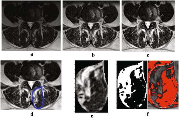
Methods: Magnetic resonance images of 30 patients with LBP were randomly selected cohort of patients participating in a project on commonly diagnosed lumbar pathologies in patients attending spine surgeon clinics. A series of T2-weighted MR images were used to train the algorithm; preprocessing techniques including adaptive histogram equalization method image adjustment scheme were used to enhance the quality and contrast of the images. All muscle measurements were repeated twice using a manual thresholding technique and the novel automated thresholding algorithm, from axial T2-weigthed images, at least 5 days apart. The rater was blinded to all earlier measurements. Inter-method agreement and intra-rater reliability for each measurement method were assessed. The study did not received external funding and the authors have no disclosures.
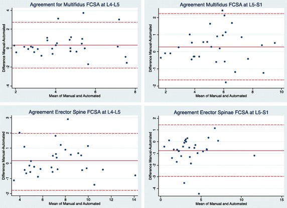
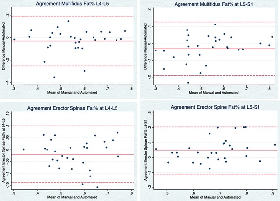
Results: There was excellent agreement between the two methods with inter-method reliability coefficients (intraclass correlation coefficients) varying from 0.79 to 0.99. Bland and Altman plots further confirmed the agreement between the two methods. Intra-rater reliability and standard error of measurements were comparable between methods, with reliability coefficient varying between 0.95 and 0.99 for the manual thresholding and 0.97-0.99 for the automated algorithm.
Conclusion: The proposed automated thresholding algorithm to assess paraspinal muscle size and composition measurements was highly reliable, with excellent agreement with the reference manual thresholding method.
Keywords: Automated algorithm; Erector spinae; Fatty infiltration; Magnetic resonance imaging; Multifidus; Paraspinal muscle.
Figures
References
-
- Kulig K, Scheid AR, Beauregard R, Popovich JM, Jr, Beneck GJ, Colletti PM. Multifidus morphology in persons scheduled for single-level lumbar microdiscectomy: qualitative and quantitative assessment with anatomical correlates. Am J Phys Med Rehabil. 2009;88(5):355–361. doi: 10.1097/PHM.0b013e31819c506d. - DOI - PubMed
Publication types
MeSH terms
LinkOut - more resources
Full Text Sources
Other Literature Sources
Medical
Miscellaneous
