. 2017 Jul 7;292(27):11218-11229.
doi: 10.1074/jbc.M117.786269. Epub 2017 May 22.

Structure-based prediction of Wnt binding affinities for Frizzled-type cysteine-rich domains

Affiliations

Structure-based prediction of Wnt binding affinities for Frizzled-type cysteine-rich domains

Mark Agostino et al. J Biol Chem. .

Abstract

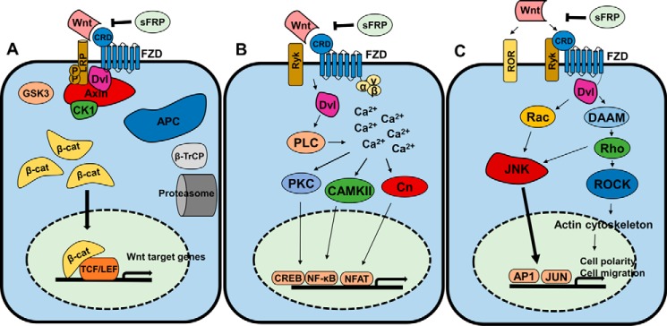
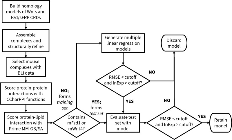
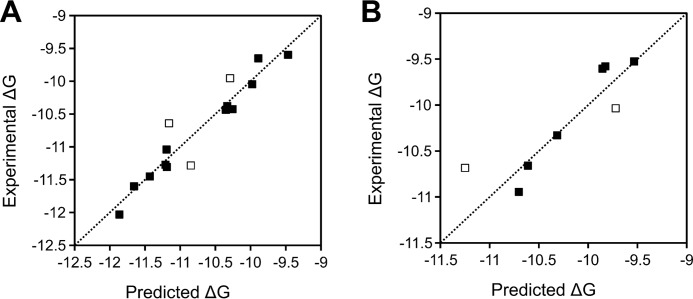
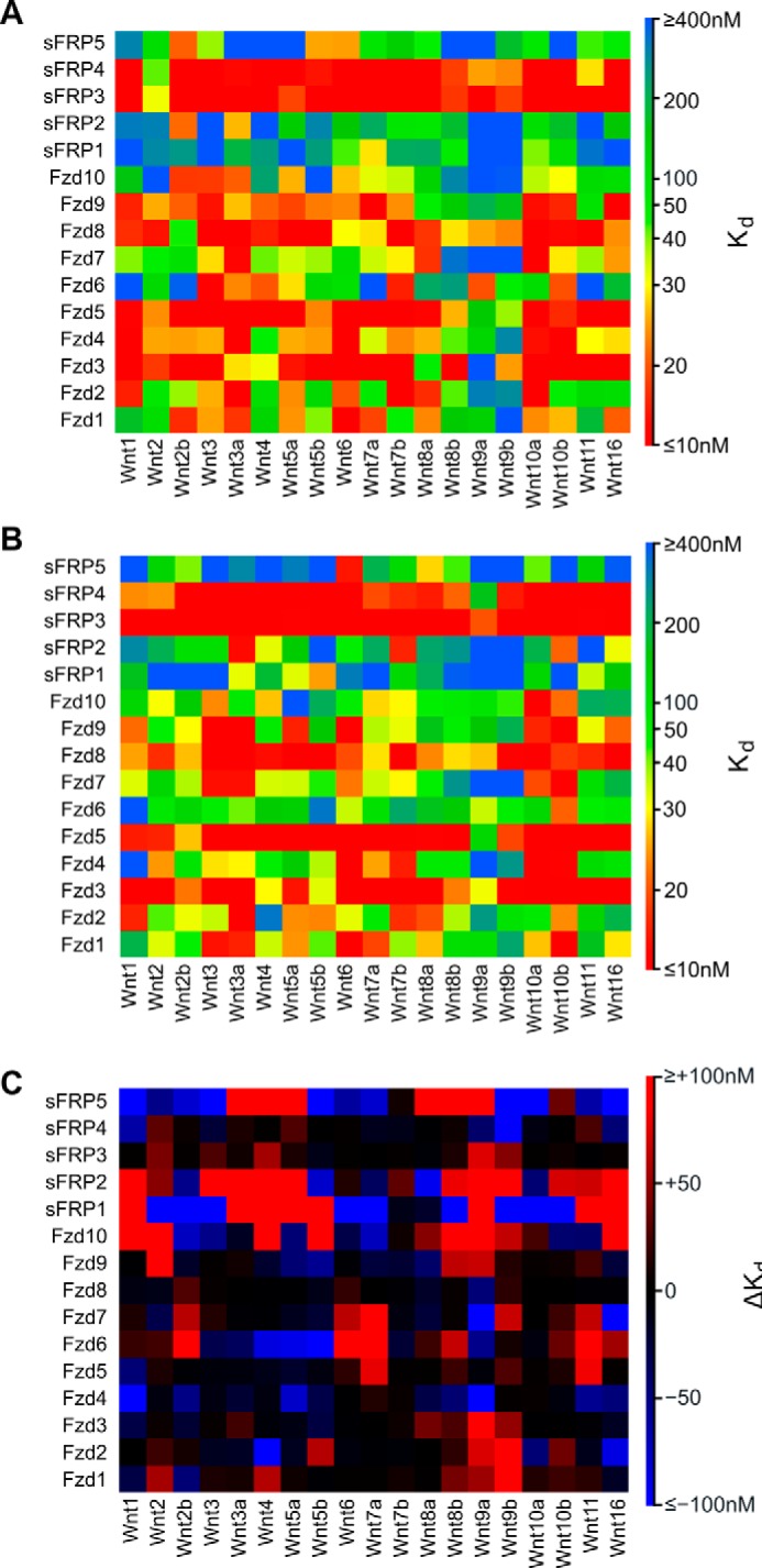
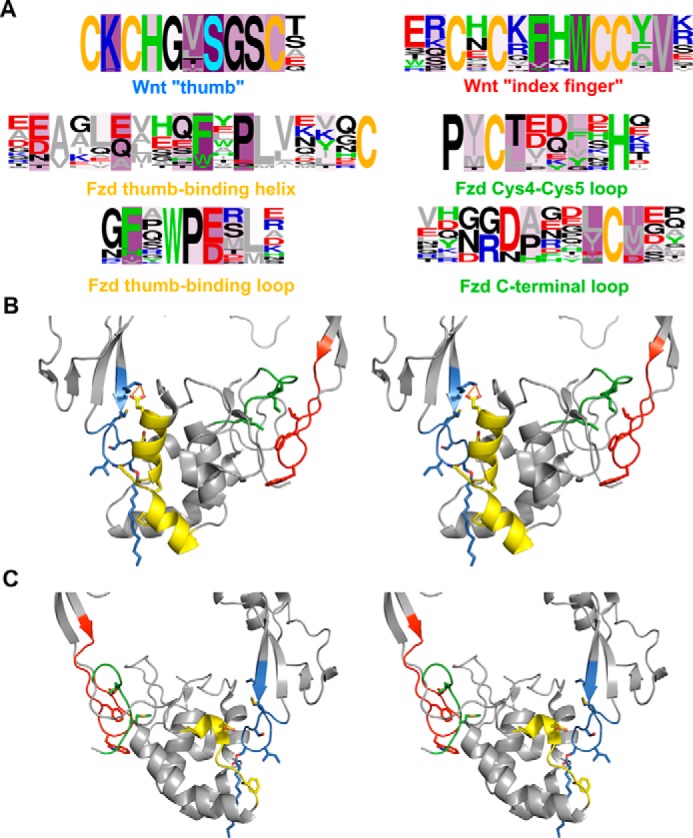
Wnt signaling pathways are of significant interest in development and oncogenesis. The first step in these pathways typically involves the binding of a Wnt protein to the cysteine-rich domain (CRD) of a Frizzled receptor. Wnt-Frizzled interactions can be antagonized by secreted Frizzled-related proteins (SFRPs), which also contain a Frizzled-like CRD. The large number of Wnts, Frizzleds, and SFRPs, as well as the hydrophobic nature of Wnt, poses challenges to laboratory-based investigations of interactions involving Wnt. Here, utilizing structural knowledge of a representative Wnt-Frizzled CRD interaction, as well as experimentally determined binding affinities for a selection of Wnt-Frizzled CRD interactions, we generated homology models of Wnt-Frizzled CRD interactions and developed a quantitative structure-activity relationship for predicting their binding affinities. The derived model incorporates a small selection of terms derived from scoring functions used in protein-protein docking, as well as an energetic term considering the contribution made by the lipid of Wnt to the Wnt-Frizzled binding affinity. Validation with an external test set suggests that the model can accurately predict binding affinity for 75% of cases and that the error associated with the predictions is comparable with the experimental error. The model was applied to predict the binding affinities of the full range of mouse and human Wnt-Frizzled and Wnt-SFRP interactions, indicating trends in Wnt binding affinity for Frizzled and SFRP CRDs. The comprehensive predictions made in this study provide the basis for laboratory-based studies of previously unexplored Wnt-Frizzled and Wnt-SFRP interactions, which, in turn, may reveal further Wnt signaling pathways.

Keywords: Frizzled receptor; Wnt signaling; cysteine-rich domain; homology modeling; lipid-protein interaction; protein-protein interaction; structural biology; structure-activity relationship.


Conflict of interest statement

The authors declare that they have no conflicts of interest with the contents of this article.

Figures

Figure 1.
Figure 1.
Wnt signaling pathways. A, canonical Wnt signaling. Wnt binding to Fzd CRD initiates the destabilization of the cytoplasmic destruction complex (adenomatous polyposis coli protein (APC), Axin, GSK3, CK1, and Dvl). This allows cytosolic β-catenin (β-cat) accumulation and subsequent translocation to the nucleus where it binds to T cell factor/lymphoid enhancer-binding factor (TCF/LEF) transcription factors to transcribe Wnt target genes. SFRPs antagonize this cascade, and β-catenin is polyubiquitinated by β-transducin repeats-containing protein (β-TrCP) and degraded by proteolysis. B, the Wnt/Ca2+ pathway. Wnt binding to Fzd CRD or Ryk co-receptor activates Dvl, which stimulates calcium release. Downstream effectors PKC, calmodulin-dependent protein kinase II (CaMKII), and Cn activate transcription factors cAMP-response element-binding protein (CREB), NF-κB, and nuclear factor of activated T cells (NFAT). C, the PCP pathway. Wnt stimulation is effected initially through Fzd-Dvl interaction and co-receptors ROR/Ryk and passed through multiple effectors (Rac, phospholipase C (PLC), Disheveled-associated activator of morphogenesis (DAAM)) downstream to ROCK and JNK. ROCK regulates the actin cytoskeleton, and JNK activates AP1 and JUN transcription factors to regulate cell polarity and migration.
Figure 2.
Figure 2.
Homology models of selected Wnt-Fzd CRD complexes overlaid to the repaired XWnt8-mFzd8 CRD crystal structure (Protein Data Bank code 4F0A). Gray, repaired Protein Data Bank code 4F0A; pink, mWnt5-mFzd1 CRD complex; yellow, mWnt10a-mFzd6 complex; green, hWnt3a-hSFRP4 CRD complex; cyan, hWnt2b-hFzd9 CRD complex. Lipid is shown in all structure as sticks with transparent spheres.
Figure 3.
Figure 3.
Overview of the model building process. RMSE and InExp cutoffs used to select models at the relevant stages of model building are described in the Experimental Procedures.
Figure 4.
Figure 4.
Comparison of binding energy predictions by Model 1 in the training set (A) and test set (B). Points indicated by open squares are those where the predicted binding energy falls outside the model RMSE (0.23 kcal/mol for the training set; 0.27 kcal/mol for the test set).
Figure 5.
Figure 5.
Binding affinity predictions by Model 1 for Wnt-Fzd interactions. A, mouse interactions. B, human interactions. C, binding affinity differences (ΔKd) between equivalent Wnt-Fzd interactions of mouse and human calculated as ΔKd = mouse Kd − human Kd. Positive ΔKd is indicative of a lower affinity interaction in mouse compared with human; negative ΔKd indicates a higher affinity interaction in mouse compared with human.
Figure 6.
Figure 6.
Residues making significant contributions to the binding energy in the majority of Wnt-Fzd CRD complexes. A, logo analyses of Wnts (first row) and Fzd CRDs (second two rows) highlighting the major regions involved in interactions. Fzd sequences interacting with a specific region of Wnt are shown below the sequence of Wnt corresponding to that region. Green, aromatic residues (Trp, Phe, His, and Tyr); gray, aliphatic residues (Val, Leu, Ile, Met, and Ala); blue, basic residues (Arg and Lys); red, acidic residues (Asp and Glu); yellow, cysteine; light blue, palmitoleoylserine (A only); black, all other residues (Gly, Pro, Ser, Thr, Gln, and Asn). Logos are presented as frequency plots. Intensity of purple shading indicates the number of complexes in which the residue at that position is a significant contributor to the complex binding energy. B and C, cross-eyed stereoviews of the “front” (B) and “rear” (C) of the XWnt8-mFzd8 crystal structure complex (Protein Data Bank code 4F0A) with major interacting regions highlighted. Residues corresponding to positions frequently involved in interactions across the full set of Wnt-Fzd CRD complexes are shown as sticks. Regions are colored according to the caption color in panel A. The front view displays the regions of the middle row of panel A; the rear view displays the regions of the bottom row of panel A.

Similar articles

Cited by

References

    1. Holstein T. W. (2012) The evolution of the Wnt pathway. Cold Spring Harb. Perspect. Biol. 4, a007922. - PMC - PubMed
    1. Hikasa H., and Sokol S. Y. (2013) Wnt signaling in vertebrate axis specification. Cold Spring Harb. Perspect. Biol. 5, a007955. - PMC - PubMed
    1. Polakis P. (2012) Wnt signaling in cancer. Cold Spring Harb. Perspect. Biol. 4, a008052. - PMC - PubMed
    1. Komiya Y., and Habas R. (2008) Wnt signal transduction pathways. Organogenesis 4, 68–75 - PMC - PubMed
    1. Pohl S. Ö.-G, Brook N., Agostino M., Arfuso F., Kumar A. P., and Dharmarajan A. (2017) Wnt signaling in triple-negative breast cancer. Oncogenesis 6, e310. - PMC - PubMed

Publication types

Associated data