. 2017 Jun 6;114(23):E4574-E4581.
doi: 10.1073/pnas.1703057114. Epub 2017 May 22.

Selective regulation of Notch ligands during angiogenesis is mediated by vimentin

Affiliations

Selective regulation of Notch ligands during angiogenesis is mediated by vimentin

Daniel Antfolk et al. Proc Natl Acad Sci U S A. .

Abstract

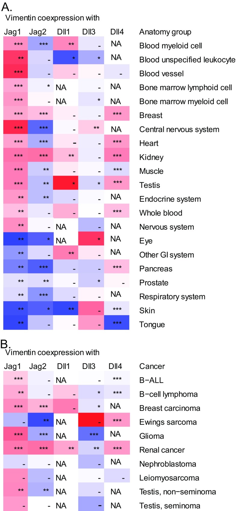
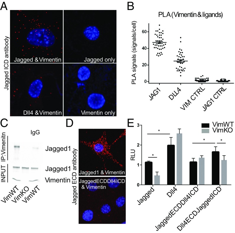
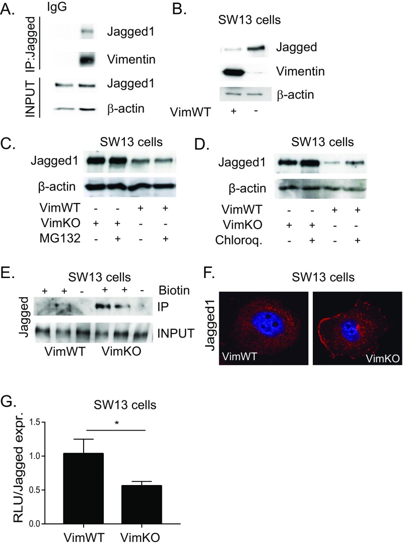
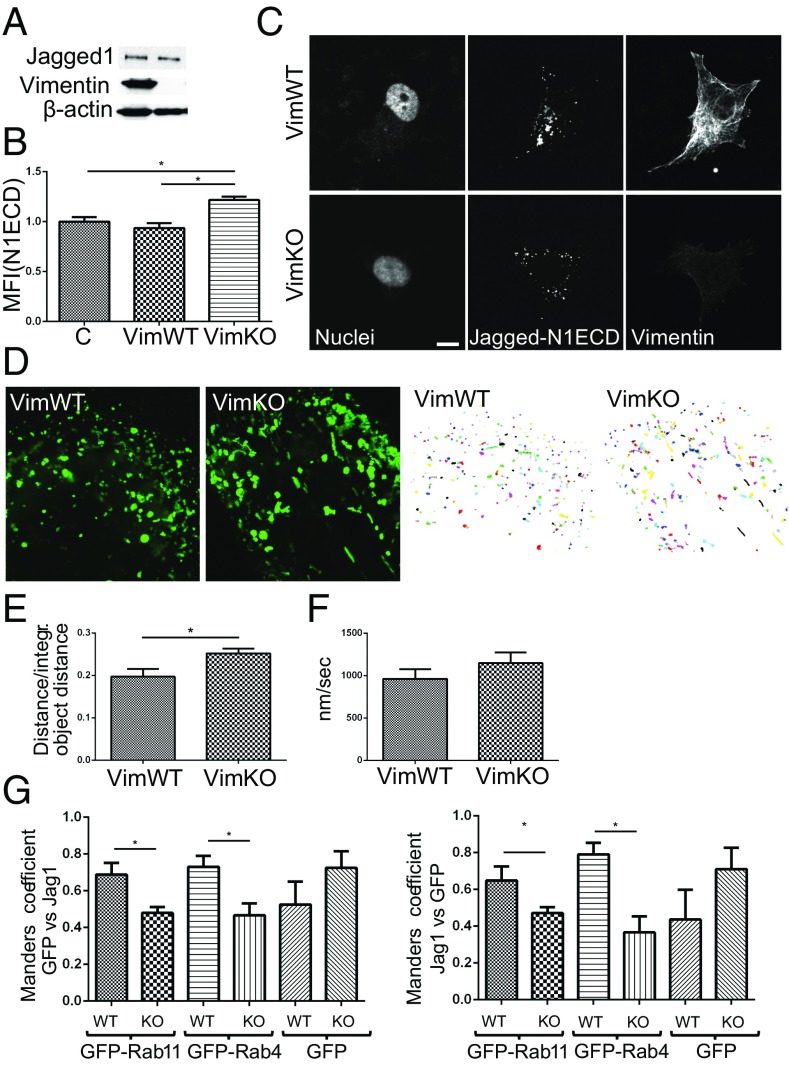
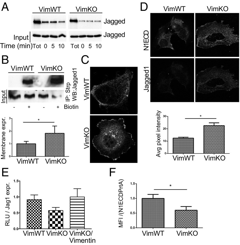
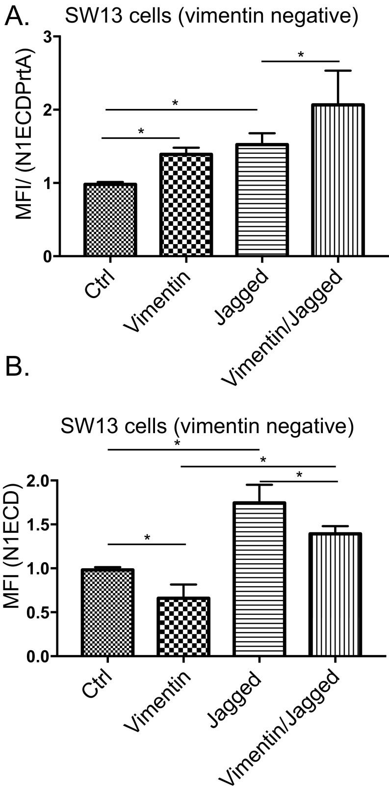
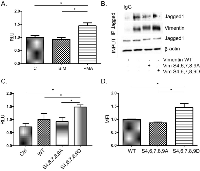
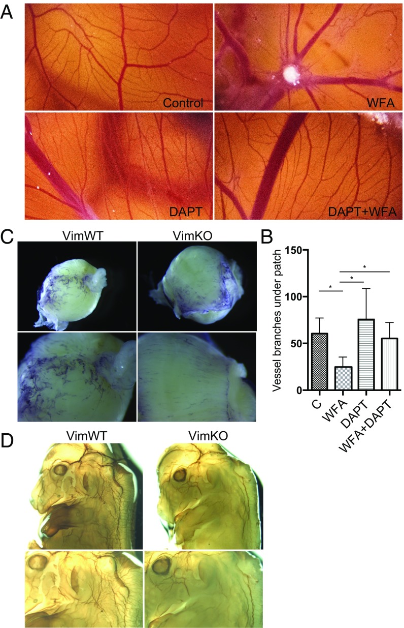
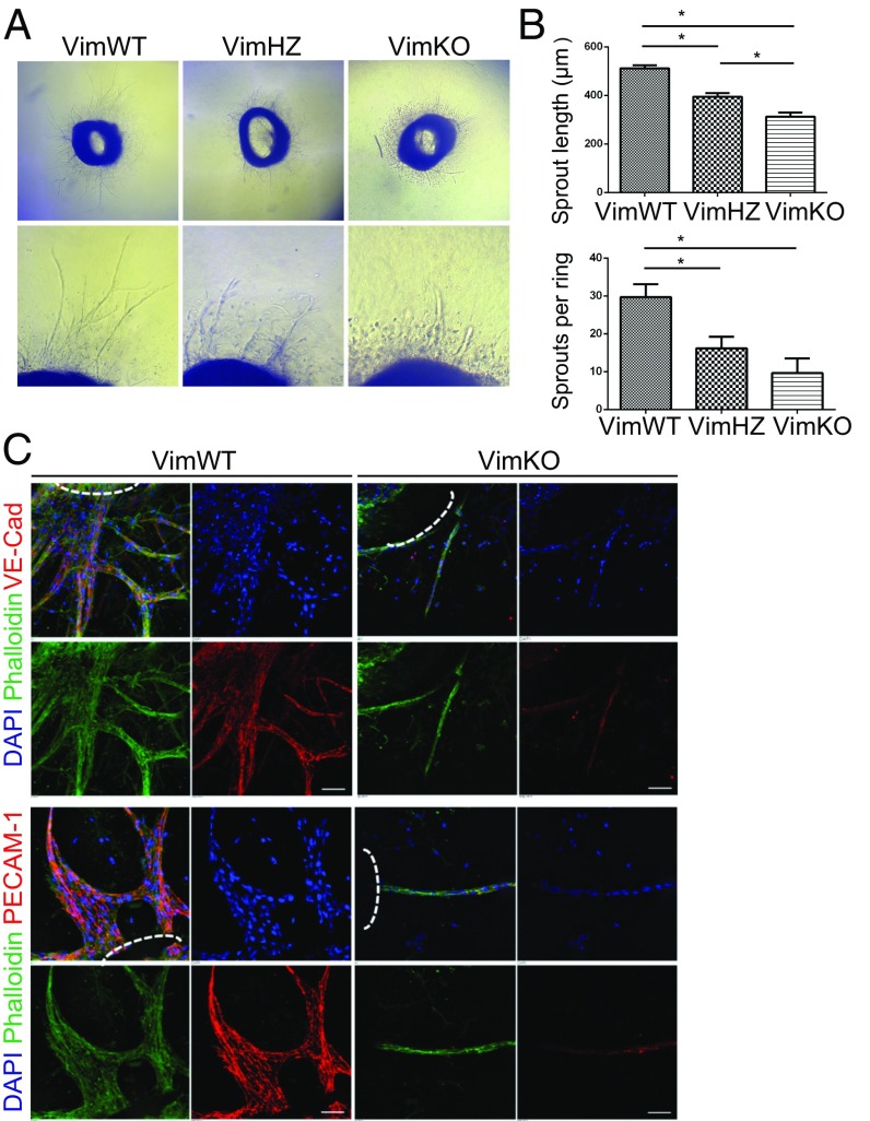
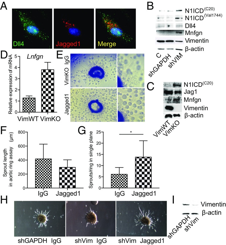
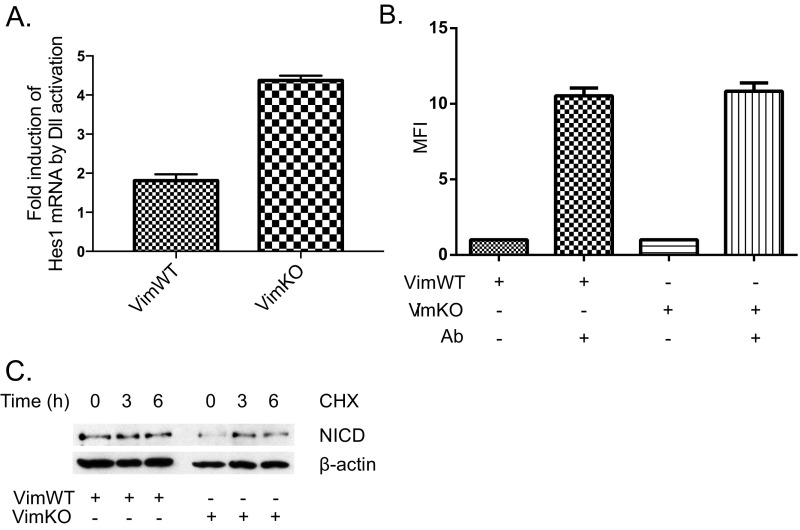
Notch signaling is a key regulator of angiogenesis, in which sprouting is regulated by an equilibrium between inhibitory Dll4-Notch signaling and promoting Jagged-Notch signaling. Whereas Fringe proteins modify Notch receptors and strengthen their activation by Dll4 ligands, other mechanisms balancing Jagged and Dll4 signaling are yet to be described. The intermediate filament protein vimentin, which has been previously shown to affect vascular integrity and regenerative signaling, is here shown to regulate ligand-specific Notch signaling. Vimentin interacts with Jagged, impedes basal recycling endocytosis of ligands, but is required for efficient receptor ligand transendocytosis and Notch activation upon receptor binding. Analyses of Notch signal activation by using chimeric ligands with swapped intracellular domains (ICDs), demonstrated that the Jagged ICD binds to vimentin and contributes to signaling strength. Vimentin also suppresses expression of Fringe proteins, whereas depletion of vimentin enhances Fringe levels to promote Dll4 signaling. In line with these data, the vasculature in vimentin knockout (VimKO) embryos and placental tissue is underdeveloped with reduced branching. Disrupted angiogenesis in aortic rings from VimKO mice and in endothelial 3D sprouting assays can be rescued by reactivating Notch signaling by recombinant Jagged ligands. Taken together, we reveal a function of vimentin and demonstrate that vimentin regulates Notch ligand signaling activities during angiogenesis.

Keywords: Jagged; Notch; angiogenesis; vimentin.


Conflict of interest statement

The authors declare no conflict of interest.

Figures

Fig. S1.
Fig. S1.
Vimentin and Jagged 1 correlate positively in several tissues (A) and cancers (B). mRNA correlations between vimentin and Jagged 1 (Jag1), Jagged 2 (Jag2), Delta-like 1 (Dll1), Delta-like 3 (Dll3), and Delta-like 4 (Dll4) were extracted from the GeneSapiens database. Red colors indicate a more positive correlation and blue, a more negative correlation. *P < 0.05, **P < 0.01, ***P < 0.001, ****P < 0.0001.
Fig. 1.
Fig. 1.
Jagged interacts with vimentin and the intracellular domain of Jagged potentiates signal activation. (A) In situ PLA was used to demonstrate physical interaction between vimentin and Jagged versus Dll4. Vimentin or Jagged were used as negative controls. (B) Number of PLA signals per cell. Quantification of signals is shown from three separate experiments. P values are <0.0001 for all groups compared with Jagged. (C) Jagged coimmunoprecipitates with vimentin from VimWT MEFs. (D) PLA using an antibody against the Jagged 1 extracellular domain shows strong PLA interaction between Jagged and vimentin, but the chimeric ligand JaggedECDDll4ICD where the intracellular domain of Jagged is switched to Dll4 shows no visible interaction. (E) The signal sending potential of Dll4, Jagged, and chimeric ligands, JaggedECDDll4ICD and Dll4ECDJaggedICD, was measured by coculturing ligand expressing VimKO and VimWT MEF cells with 293HEK cells expressing the Notch 1 receptor (293HEK-FLN1) using a luciferase-based reporter system. Values represent means of three separate experiments including three experimental repetitions ± SEM. Statistical significance was determined using one-way ANOVA and Bonferroni post hoc test, P < 0.05. RLU, relative light unit.
Fig. S2.
Fig. S2.
Vimentin does not modulate Jagged turnover. (A) Vimentin coimmunoprecipitates with Jagged in 293 HEK cells transfected with vimentin. (B) Representative immunoblot shows Jagged 1 protein levels in vimentin-negative and -positive SW13 cells. (C) Representative images showing Jagged 1 protein expression in vimentin-negative and -positive SW13 cells in the presence of the proteosome inhibitor MG132 (20 µM, 6 h). (D) Representative immunoblot showing Jagged 1 protein expression in vimentin-negative and -positive SW13 cells in the presence of the lysosomal inhibitor chloroquine (20 µM, 16 h). (E) Cell surface proteins in SW13 cells were labeled with biotin and immunoprecipitated using streptavidin agarose beads. Immunoblotting was performed with an antibody detecting Jagged 1. (F) Representative confocal microscopy images showing Jagged 1 immunofluorescence in vimentin-negative and -positive SW13 cells. (G) Jagged signal sending potential measured by coculturing vimentin-negative and -positive SW13 cells with 293HEK cells expressing the Notch 1 receptor (293HEK-FLN1) using a luciferase-based reporter system. Jagged signal sending potential as related to surface levels of Jagged. Values represent means ± SEM. Statistical significance was determined using Student’s t test, P < 0.05. RLU, relative light unit.
Fig. 2.
Fig. 2.
Vimentin alters Jagged 1 trafficking. (A) Representative immunoblot shows Jagged 1 protein levels in VimWT and VimKO MEFs. (B) Quantification of ligand endocytosis VimWT and VimKO cells using fluorescently labeled recombinant peptides mimicking the Notch 1 extracellular domain conjugated to a fluorescent probe (N1ECD) to label the ligands. Values represent means ± SEM. Statistical significance was determined using one-way ANOVA and Bonferroni post hoc test, *P < 0.05. MFI, mean fluorescence intensity. (C) Representative images of N1ECD localization in VimWT and VimKO cells. (Scale bar, 10 µm.) (D) VimWT and VimKO cells were live labeled with N1ECD–Alexa-488 and vesicles with green fluorescence were followed for 1 min. Projection of N1ECD–Alexa-488 positive vesicle movement in VimWT and VimKO MEFs during 1 min of vesicle tracking. Vesicles were segmented and tracked with CellProfiler. Graphs were created using R from information produced with CellProfiler. One color represents one vesicle. (E) Analysis of N1ECD–Alexa-488 labeled vesicle track linearity in VimWT and VimKO cells. Values represent means ± SEM. Statistical significance was determined using Student’s t test, P < 0.05. (F) Analysis of N1ECD–Alexa-488 labeled vesicle speed in VimWT and VimKO cells. (Scale bar, 10 µm.) (G) Ligand localization into different endosomal compartments was analyzed by evaluating the colocalization between Jagged 1-stained vesicles and Rab4- or Rab11-linked recycling endosomes. Jagged 1 colocalization with Rab4 and Rab11 was analyzed by Manders’ colocalization coefficient. Confocal microscopy images of 30 cells from three experiments described above were quantified using Fiji ImageJ. P < 0.05.
Fig. 3.
Fig. 3.
Vimentin regulates Jagged recycling and Notch activation. (A) Analysis of Jagged 1 recycling in VimWT and VimKO cells using a biotin cell surface labeling and stripping protocol. (B) Cell surface proteins were labeled with biotin and immunoprecipitated using streptavidin agarose beads. Immunoblotting was performed with an antibody detecting Jagged 1. (C) Representative confocal microscopy images showing Jagged 1 immunofluorescence in VimWT and VimKO cells. (D) Representative confocal microscopy image shows Jagged 1 cell surface localization and N1ECD binding in VimWT and VimKO cells. (Scale bar, 10 µm.) The graph shows quantification of pixel intensity. Values represent means ± SEM. Statistical significance was determined using Student’s t test, P < 0.05. (E) Jagged signal sending potential measured by coculturing VimWT and VimKO cells and VimKO cells reexpressing vimentin with 293HEK cells expressing the Notch 1 receptor (293HEK-FLN1) using a luciferase-based reporter system. Jagged signal sending potential as related to surface levels of Jagged. Values represent means ± SEM. Statistical significance was determined using Student’s t test, P < 0.05. RLU, relative light unit. (F) The ability of VimKO and VimWT to internalize N1ECD–Alexa-488 coupled to protein A agarose beads (N1ECDPrtA). Internalization of N1ECDPrtA in VimKO and VimWT cells as related to surface levels of Jagged. Values represent means ± SEM. Statistical significance was determined using Student’s t test, P < 0.05. MFI, mean fluorescence intensity.
Fig. S3.
Fig. S3.
Vimentin regulates Jagged endocytosis, Notch receptor transendocytosis, and Notch activity in vimentin-negative fibroblasts and epithelial breast cancer cells. (A) The ability of vimentin-negative control SW13 cells or cells transfected with vimentin, Jagged, or vimentin and Jagged to internalize N1ECD–Alexa-488 coupled to protein A agarose beads (N1ECDPrtA). (B) Endocytosis of Jagged in vimentin-negative SW13 cells or in cells transfected with vimentin, Jagged, or vimentin together with Jagged as measured by a fluorescently labeled N1ECD peptide. Values represent means ± SEM. Statistical significance was determined using Student's t test, P < 0.05. Values represent means ± SEM. Statistical significance was determined using Student’s t test, P < 0.05. MFI, mean fluorescence intensity.
Fig. S4.
Fig. S4.
Vimentin-mediated regulation of Jagged is vimentin phosphorylation dependent. (A) Analysis of Notch signaling potential after pretreatment with the PKC inhibitor bis-indolylmaleimide I (BIM) or the PKC activator Phorbol-12-Myristate-13-Acetate (PMA). The ability of the treated cells to activate Notch in cocultured 293 FLN cells was measured using a luciferase-based reporter system (12xCSL-luc). Values represent means ± SEM. Statistical significance was determined using one-way ANOVA and Bonferroni post hoc test, P < 0.05. (B) Immunoprecipitation of Jagged from vimentin-negative SW13 cells expressing wild-type vimentin, phosphomimicking (VimS4,6,7,8,9D), or phosphodeficient (VimS4,6,7,8,9A) forms of vimentin. (C) Notch activation potential of vimentin-negative control SW13 cells or cells transfected with wild type, phosphomimicking (VimS4,6,7,8,9D), or phosphodeficient (VimS4,6,7,8,9A). Values represent means ± SEM. Statistical significance was determined using Student’s t test, P < 0.05. (D) Receptor-transendocytosis of vimentin-negative control SW13 cells or cells transfected with wild type, phosphomimicking (VimS4,6,7,8,9D), or phosphodeficient (VimS4,6,7,8,9A). Values represent means ± SEM. Statistical significance was determined using Student’s t test, P < 0.05.
Fig. 4.
Fig. 4.
Loss of vimentin disrupts angiogenesis. (A) Notch inhibition rescues the antiangiogenic effect of WFA. The effects of the vimentin-targeted drug WFA, the γ-secretase inhibitor DAPT, and the combination of DAPT and WFA were analyzed in the CAM angiogenesis model. (B) Number of vessel branches per field of view under the patch containing the drugs in one representative experiment. (C) Whole-mount PECAM-1 immunostaining of E11.5 placental tissue from VimWT and VimKO mice. (D) Whole-mount PECAM-1 immunostaining of VimWT and VimKO embryos at E12.5.
Fig. 5.
Fig. 5.
Decreased sprouting from aortic rings from vimentin knockout mice. (A) Representative images showing endothelial sprouting in aortic rings from VimWT, VimHZ, and VimKO mice. (B) Quantification of endothelial sprout length and sprout number. Sprout length was quantified as the distance (in micrometers) from the edge of the ring to the tip of invading endothelial structure. Data represent the average number of endothelial sprouts per ring in a single plane. Error bars represent SEM. Statistical significance was determined using one-way ANOVA and Bonferroni post hoc test, P < 0.05. (C) Immunofluorescence staining was performed to determine whether sprouting structures contained endothelial and smooth muscle cells. Aortic ring assays using VimWT and VimKO aortic rings were stained with VE-cadherin and phalloidin-FITC (Upper) or PECAM-1 and phalloidin-FITC (Lower). In all panels, overlap and DAPI images are shown in Upper Left and Right images, respectively. Z stacks of 1-µm step-size compressed images of aortic rings were taken using a Nikon TI A1R inverted confocal microscope at 40× magnification. (Scale bar, 100 μm.) Dotted white line represents edge of aortic ring where outgrowth initiates.
Fig. S5.
Fig. S5.
Aortic ring assay. To determine the most appropriate conditions during aortic ring assays for robust endothelial outgrowth, aortic rings were fed Opti-MEM containing 2.5% FBS supplemented with nothing (control, C), 40 ng/mL VEGF, 40 ng/mL FGF, or EGM2-MV.
Fig. 6.
Fig. 6.
Immobilized Notch Jagged 1 rescues sprouting. (A) Confocal image showing Dll4 and Jagged 1 immunolabeling in HUVECs. (B) Expression of Notch receptors, ligands, and regulators in control or vimentin knockdown HUVECs as determined by immunoblotting. The immunoblot shows expression of NICD by two different antibodies: C20 and Val1744, and Dll4, Manic Fringe (Mnfgn), and vimentin. β-Actin was used as a loading control. (C) Expression of Notch receptors, ligands, and regulators in endothelial cells isolated from VimWT or VimKO mice as determined by immunoblotting. Images from B and C are composites, and the bands were taken from several gels run from the same samples with equal loading. (D) The influence of vimentin on the expression of Lunatic Fringe determined by qPCR. (E) To determine whether exogenous addition of Notch ligands rescued vimentin null sprouting responses, Notch ligands or IgG control were coupled to protein A agarose beads and mixed with collagen before embedding vimentin null aortic rings. Representative images show endothelial sprouting in aortic rings from VimKO mice in the presence of immobilized Notch ligands Dll4 and Jagged1. (F) Quantification of endothelial sprout length in aortic rings from VimKO mice in the presence of IgG or immobilized Notch ligands Dll4 and Jagged 1. Sprout length was quantified as the distance (in micrometers) from the edge of the ring to the tip of the invading endothelial structure. (G) Quantification of the number of endothelial sprouts per ring in a single plane. Values represent means ± SEM. Statistical significance was determined using Student’s t test, P < 0.05. (H) Representative images of an in vitro 3D angiogenesis assay with HUVECs transfected with shRNA against vimentin. Endothelial sprouting was analyzed in the presence of IgG or immobilized Notch ligands Dll4 and Jagged 1. (I) Expression of vimentin in control or vimentin knockdown HUVECs as determined by immunoblotting.
Fig. S6.
Fig. S6.
Dll-mediated Notch activation is enhanced in VimKO cells. (A) Levels of Notch receptors on the surface of VimWT and VimKO cells were analyzed by flow cytometry (FACS) using an antibody recognizing the extracellular domain of Notch. Cells were labeled at +4 °C, before harvesting and analyzes by FACS. (B) VimWT and VimKO cells were cultured on Dll-FC ligands immobilized to G protein beads for 6 h (58). The cells were harvested, mRNA isolated, and samples subsequently analyzed for expression of the Notch target gene Hes1. The graph denotes fold induction of Hes1 expression in VimWT and VimKO cells as related to expression in cells grown on FC immobilized to protein G beads (58). (C) Cyclohexamide treatment of VimWT and VimKO cells demonstrated that NICD in VimWT and VimKO remains stable for 6 h, excluding an effect on Hes1 expression by differences in NICD turnover in WT and KO cells.

References

    1. Schiffers PM, et al. Altered flow-induced arterial remodeling in vimentin-deficient mice. Arterioscler Thromb Vasc Biol. 2000;20:611–616. - PubMed
    1. Kwak HI, et al. Calpain-mediated vimentin cleavage occurs upstream of MT1-MMP membrane translocation to facilitate endothelial sprout initiation. Angiogenesis. 2012;15:287–303. - PMC - PubMed
    1. Ivaska J, et al. PKCepsilon-mediated phosphorylation of vimentin controls integrin recycling and motility. EMBO J. 2005;24:3834–3845. - PMC - PubMed
    1. Nieminen M, et al. Vimentin function in lymphocyte adhesion and transcellular migration. Nat Cell Biol. 2006;8:156–162. - PubMed
    1. Pallari HM, Eriksson JE. Intermediate filaments as signaling platforms. Sci STKE. 2006;2006:pe53. - PubMed

Publication types

MeSH terms