. 2017 Jun 6;114(23):5994-5999.
doi: 10.1073/pnas.1703546114. Epub 2017 May 22.

Insect pathogenic fungus interacts with the gut microbiota to accelerate mosquito mortality

Affiliations

Insect pathogenic fungus interacts with the gut microbiota to accelerate mosquito mortality

Ge Wei et al. Proc Natl Acad Sci U S A. .

Abstract

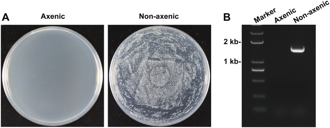
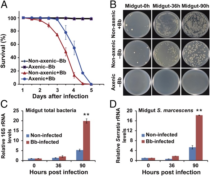
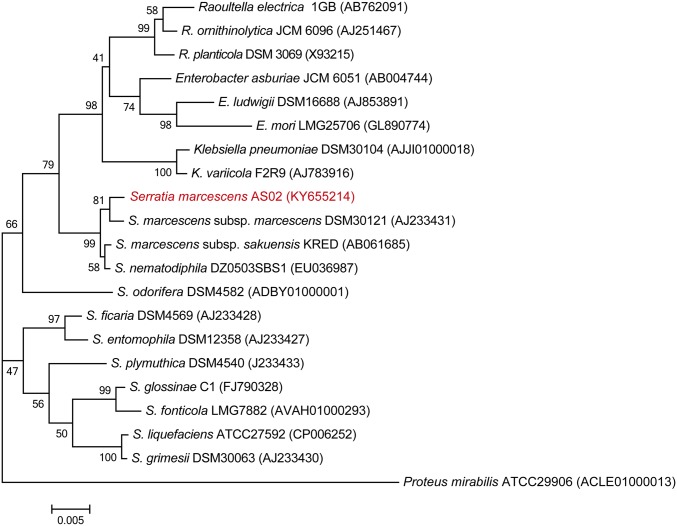
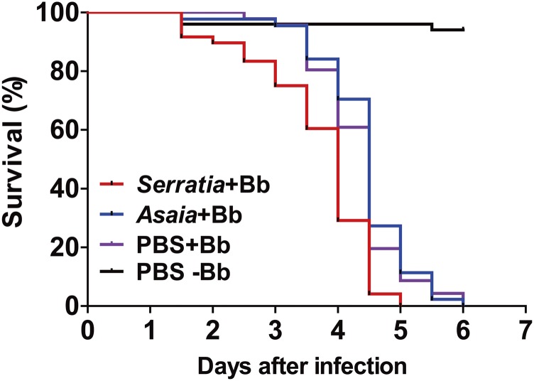
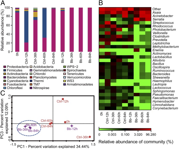
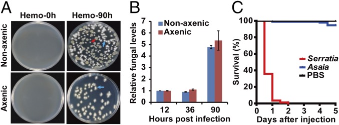
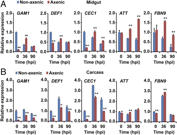
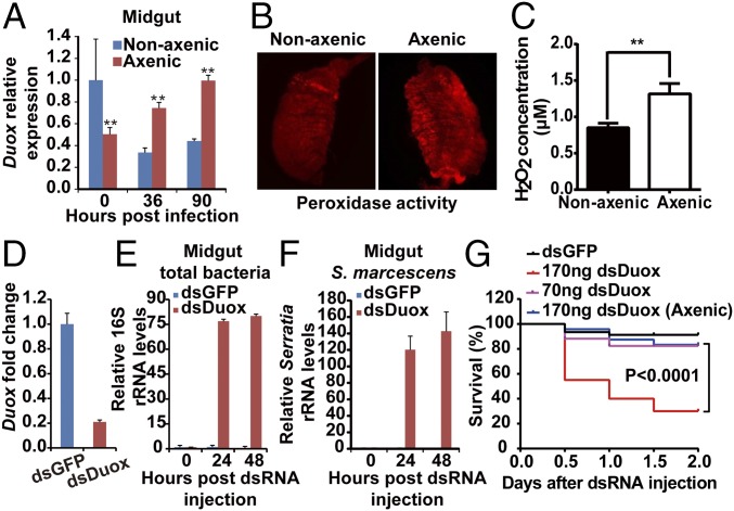
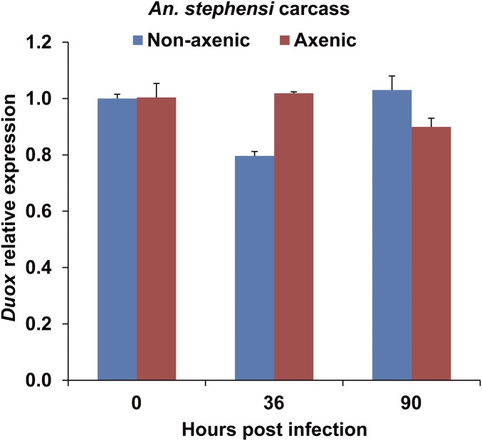
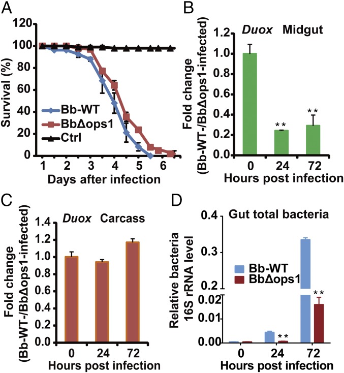
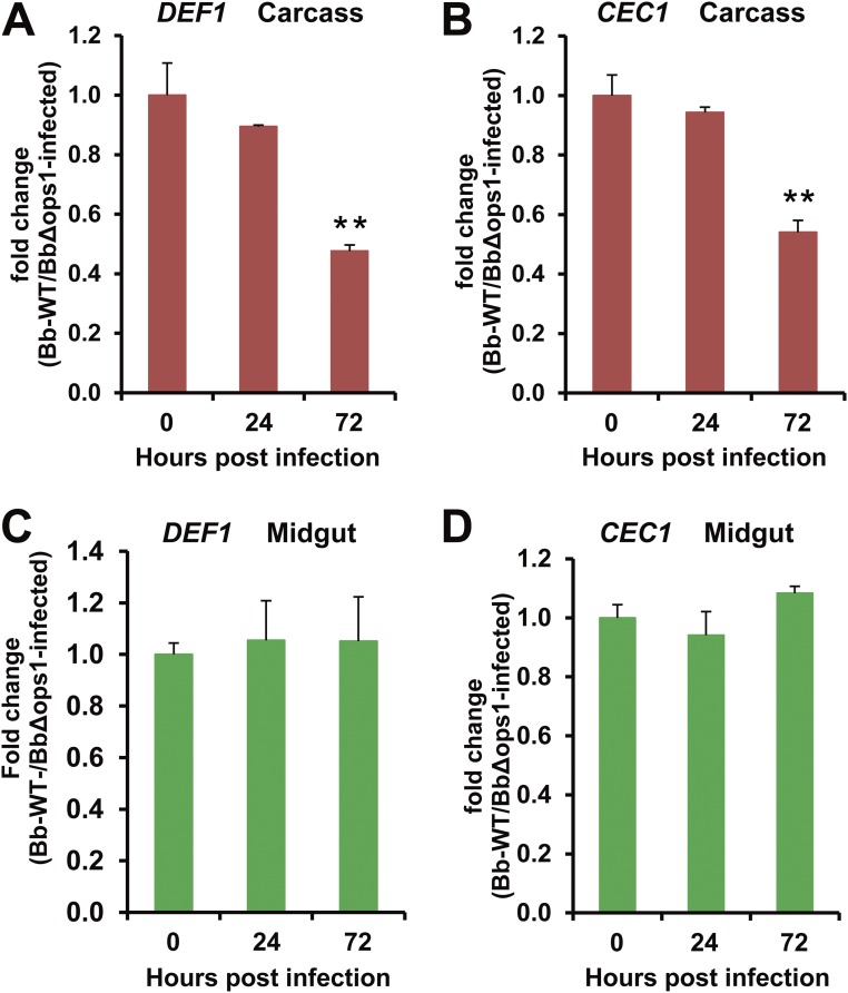
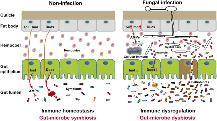
The insect gut microbiota plays crucial roles in modulating the interactions between the host and intestinal pathogens. Unlike viruses, bacteria, and parasites, which need to be ingested to cause disease, entomopathogenic fungi infect insects through the cuticle and proliferate in the hemolymph. However, interactions between the gut microbiota and entomopathogenic fungi are unknown. Here we show that the pathogenic fungus Beauveria bassiana interacts with the gut microbiota to accelerate mosquito death. After topical fungal infection, mosquitoes with gut microbiota die significantly faster than mosquitoes without microbiota. Furthermore, fungal infection causes dysbiosis of mosquito gut microbiota with a significant increase in gut bacterial load and a significant decrease in bacterial diversity. In particular, the opportunistic pathogenic bacterium Serratia marcescens overgrows in the midgut and translocates to the hemocoel, which promotes fungal killing of mosquitoes. We further reveal that fungal infection down-regulates antimicrobial peptide and dual oxidase expression in the midgut. Duox down-regulation in the midgut is mediated by secretion of the toxin oosporein from B. bassiana Our findings reveal the important contribution of the gut microbiota in B. bassiana-killing activity, providing new insights into the mechanisms of fungal pathogenesis in insects.

Keywords: Anopheles; dysbiosis; entomopathogenic fungus; gut microbiota; immunity.


Conflict of interest statement

The authors declare no conflict of interest.

Figures

Fig. S1.
Fig. S1.
Generation of axenic mosquitoes. The efficacy of elimination of midgut bacteria confirmed by (A) culturing mosquito (n = 10) gut homogenates on LB agar plates and (B) by performing PCR analysis on mosquito (n = 10) gut homogenates using universal 16S rRNA gene primers.
Fig. 1.
Fig. 1.
Effect of gut microbiota on pathogenesis in A. stephensi infected by the fungus B. bassiana (Bb). (A) Survival of axenic (without microbiota) and nonaxenic (with microbiota) mosquitoes (n = 50) following topical infection (+Bb) or no topical infection (−Bb) with B. bassiana. (B) Load of midgut cultivable bacteria from Bb-infected nonaxenic, noninfected nonaxenic, and noninfected axenic mosquitoes (n = 20) at 0, 36, and 90 h post fungal infection. Bacterial load was determined by plating the homogenate of mosquito midguts with 10,000 dilution on LB agar plates. Representative images are shown. (C and D) Number of midgut total bacteria (C) and S. marcescens (D) in Bb-infected and noninfected mosquitoes (n = 15) at 0, 36, and 90 hpi; quantification was by 16S rRNA gene-based qPCR analysis. Three biological replicates were conducted. Error bars indicate SD. Double asterisks represent a significant difference determined by the Student’s t test at P < 0.01.
Fig. S2.
Fig. S2.
Phylogenetic analysis of the translocating isolate S. marcescens AS02 with 20 related bacteria. The phylogenetic relationships were inferred from the alignment of 1,508 bp of 16S rRNA gene indicating the positions of S. marcescens AS02 relative to selected Serratia species and other genera within the family Enterobacteriaceae. The evolutionary history was inferred by using the neighbor-joining method conducted in MEGA7 software. A sequence of Proteus mirabilis (a member of the Enterobacteriaceae family) was used as the outgroup. Bootstrap values (percentages of 1,000 tree replications) greater than 40% are displayed at the nodes. Sequence GenBank accession numbers are shown in parentheses. (Bar, 0.005 substitutions per nucleotide position.)
Fig. S3.
Fig. S3.
Survival of mosquitoes (n = 50) fed S. marcescens or Asaia sp. after topical infection with a 5 × 10⁸ conidia per mL suspension of B. bassiana. Control mosquitoes were fed PBS. Bacteria were introduced into adult mosquito midgut via a sugar meal; female adults were allowed to feed for 24 h on 5% sucrose containing bacteria at a final concentration of 108 cells per mL. Introduction of S. marcescens (Serratia+Bb) significantly increased mosquito susceptibility to fungal infection compared with the PBS+Bb treatment [log-rank (Mantel–Cox) test, P < 0.001]. The experiments were performed in three biological replicates. The log-rank test was used to assess the significance of differences between two survival curves using GraphPad Prism software.
Fig. 2.
Fig. 2.
Fungal infection alters the composition of gut microbiota in mosquitoes. (A) Histogram showing temporal changes, at the phylum level, in noninfected (Ctrl; Triton treatment as control) and Bb-infected mosquitoes (n = 40) over 84 h. (B) Heat map showing temporal changes, at the genus level, in Ctrl and Bb-infected mosquitoes. (C) Principal component analysis of unweighted jack-knifed UniFrac distances of microbial communities from Ctrl and Bb-infected mosquitoes.
Fig. 3.
Fig. 3.
Translocation of gut bacteria to mosquito hemocoel after topical infection with B. bassiana. (A) Growth of bacteria and fungi from the hemolymph of nonaxenic and axenic mosquitoes at 0 and 90 hpi with B. bassiana. The red arrow indicates bacterial colonies. The blue arrows indicate fungal colonies. (B) qPCR-based quantification of fungal load in nonaxenic and axenic mosquitoes (n = 15) at 12, 36, and 90 hpi. Fungal levels are expressed as that of fungal gpd mRNA relative to A. stephensi ribosomal protein S7 (AsS7) mRNA. (C) Survival of mosquitoes (n = 100) following injection of 100 CFUs of S. marcescens, 100 CFUs of Asaia sp., or PBS (control) into the hemolymph. Experiments were performed in three replicates with similar results. Error bars indicate SD.
Fig. 4.
Fig. 4.
Expression of five AMPs in the midgut is down-regulated after topical infection with B. bassiana. qPCR analysis of expression levels of AMPs in the (A) midgut and (B) carcass of nonaxenic and axenic mosquitoes (n = 20) at 0, 36, and 90 hpi. Gene expression of each sample was normalized to that of nonaxenic mosquitoes at time 0 (taken as 1). Three biological replicates were conducted. Error bars indicate SD. Single and double asterisks represent a significant difference determined by the Student’s t test at P < 0.05 and P < 0.01, respectively.
Fig. 5.
Fig. 5.
Expression of mosquito Duox following fungal infection and effect of Duox silencing on midgut bacterial load and host survival. (A) Duox mRNA levels in the midgut of nonaxenic and axenic mosquitoes (n = 20) at 0, 36, and 90 hpi with B. bassiana. (B) Fluorescence staining for peroxidase activity. (C) H2O2 concentration in the midgut of nonaxenic and axenic mosquitoes (n = 5) infected by B. bassiana at 60 h. (D) Midgut Duox silencing efficiency in mosquitoes (n = 20) injected with 70 ng of dsGFP or dsDuox. (E and F) Effect of Duox silencing on midgut total bacterial load (E) (n = 15) and S. marcescens (F) at 0, 24, and 48 h post dsRNA injection (n = 15); levels are relative to readings at time 0 taken as 1. (G) Survival of Duox-silenced nonaxenic and axenic mosquitoes (n = 50) injected with different amounts of dsDuox. Experiments were performed in three biological replicates. Error bars indicate SD. Double asterisks represent a significant difference determined by the Student’s t test at P < 0.01.
Fig. S4.
Fig. S4.
Expression of Duox in the carcass of nonaxenic and axenic mosquitoes following fungal infection. Quantitative RT-PCR analysis of Duox mRNA levels in the carcass of nonaxenic and axenic mosquitoes (n = 20) at 0, 36, and 90 h post topical infection with B. bassiana. Error bars indicate SD. Three biological replicates were conducted.
Fig. 6.
Fig. 6.
Effect of oosporein on fungal virulence, mosquito Duox expression, and midgut bacterial growth. (A) Survival of adult female A. stephensi (n = 50) following topical infection with B. bassiana WT or BbΔops1; control mosquitoes were not infected. (B and C) Effect of Bbops1 disruption on mRNA levels of Duox in the midgut (B) (n = 20) and carcass (C) of mosquitoes (n = 20) following topical infection with B. bassiana WT and BbΔops1. (D) Effect of Bbops1 disruption on midgut total bacteria determined by 16S rRNA gene-based qPCR analysis (n = 15). Experiments were conducted in three biological replicates. Error bars indicate SD. Double asterisks represent a significant difference determined by the Student’s t test at P < 0.01.
Fig. S5.
Fig. S5.
Effect of Bbops1 disruption on mRNA expression of DEF1 and CEC1. mRNA levels of DEF1 in the (A) carcass and (C) midgut, and mRNA levels of CEC1 in the (B) carcass and (D) midgut of mosquitoes (n = 20) following topical infection with B. bassiana WT and BbΔops. Error bars indicate SD. Double asterisks represent a significant difference determined by the Student’s t test at P < 0.01. Three biological replicates were performed.
Fig. S6.
Fig. S6.
Model of the proposed mechanism of B. bassiana interactions with the gut microbiota to accelerate mosquito death. When the mosquitoes are topically infected by B. bassiana, expression of antimicrobial peptides (AMPs) and Duox in the midgut is down-regulated. Duox down-regulation in the midgut is mediated by the toxin oosporein secreted from B. bassiana. The immune dysregulation in the midgut might result in dysbiosis of gut microbiota and translocation of the opportunistic pathogenic bacterium S. marcescens from the gut to the hemocoel, where switching from asymptomatic gut symbiont to hemocoelic pathogen accelerates the killing of mosquitoes by the fungus.

References

    1. Medlock JM, et al. A review of the invasive mosquitoes in Europe: Ecology, public health risks, and control options. Vector Borne Zoonotic Dis. 2012;12:435–447. - PMC - PubMed
    1. Wang S, Jacobs-Lorena M. Genetic approaches to interfere with malaria transmission by vector mosquitoes. Trends Biotechnol. 2013;31:185–193. - PMC - PubMed
    1. Fan Y, Borovsky D, Hawkings C, Ortiz-Urquiza A, Keyhani NO. Exploiting host molecules to augment mycoinsecticide virulence. Nat Biotechnol. 2012;30:35–37. - PubMed
    1. Kanzok SM, Jacobs-Lorena M. Entomopathogenic fungi as biological insecticides to control malaria. Trends Parasitol. 2006;22:49–51. - PubMed
    1. Lord JC. From Metchnikoff to Monsanto and beyond: The path of microbial control. J Invertebr Pathol. 2005;89:19–29. - PubMed

Publication types