. 2017 Jun 6;114(23):6040-6045.
doi: 10.1073/pnas.1619843114. Epub 2017 May 22.

Nanomechanics of the substrate binding domain of Hsp70 determine its allosteric ATP-induced conformational change

Affiliations

Nanomechanics of the substrate binding domain of Hsp70 determine its allosteric ATP-induced conformational change

Soumit Sankar Mandal et al. Proc Natl Acad Sci U S A. .

Abstract

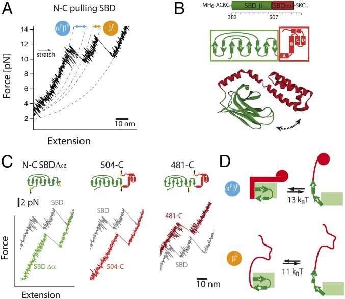
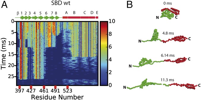
Owing to the cooperativity of protein structures, it is often almost impossible to identify independent subunits, flexible regions, or hinges simply by visual inspection of static snapshots. Here, we use single-molecule force experiments and simulations to apply tension across the substrate binding domain (SBD) of heat shock protein 70 (Hsp70) to pinpoint mechanical units and flexible hinges. The SBD consists of two nanomechanical units matching 3D structural parts, called the α- and β-subdomain. We identified a flexible region within the rigid β-subdomain that gives way under load, thus opening up the α/β interface. In exactly this region, structural changes occur in the ATP-induced opening of Hsp70 to allow substrate exchange. Our results show that the SBD's ability to undergo large conformational changes is already encoded by passive mechanics of the individual elements.

Keywords: elasticity; force; laser trapping; parallel pathways; protein extension.


Conflict of interest statement

The authors declare no conflict of interest.

Figures

Fig. 1.
Fig. 1.
Single-molecule force experiments of the SBD of the Hsp70 chaperone. (A) Three-dimensional structure of the Hsp70 chaperone DnaK in the ADP/apo state (PDB ID code 2KHO; ref. 30) and the ATP state (PDB ID code 4B9Q; ref. 8). The protein consists of two domains: the nucleotide binding domain, which is further divided into lobe I (blue) and lobe II (red), and the substrate binding domain, which is divided into the α-subdomain and the β-subdomain. The figures were prepared by using DS Viewer (Accelrys Software). (B) Optical trap assay. The SBD (green/red surfaces) is tethered to the beads (gray spheres) by two DNA handles, and beads are trapped in highly focused laser beams (red cones). The connection between the DNA and protein is realized by the modification of the two cysteine residues of the protein by the single-stranded DNA-maleimide oligonucleotide complementary to the DNA-handle overhang (for details, see ref. 16). One of the beams is reflected by a steerable mirror, which enables pulling and stretching of a single protein. The conditions were 10 mM Na2HPO4, 1.8 mM KH2PO4, 2.7 mM KCl, and 137 mM NaCl (PBS buffer), pH 7.5 at 30 °C. (C) Shown is a sequence of force-extension curves of a single SBD domain. The protein-DNA construct was stretched several times (black curves, 1 kHz filtered) at constant velocity of 200 nm/s. In every individual trace, two unfolding events of different contour-length increase are visible. The dashed lines correspond to worm-like chain (WLC) fits. (D) The order of the individual unfolding events varies. Pathway I corresponds to unfolding of the larger fragment first, followed by the shorter SBD fragment. For pathway II, the order of unfolding events is reversed. (E) Force extension curves of the SBDΔα construct at 200 nm/s (1 kHz). (F) Scatter plot of unfolding forces and corresponding contour length increases with respective distributions for pathway I and pathway II (red, α-helical part; green, β-subdomain). We found significant fluctuations at forces > 9 pN (Fig. 2A), hence the worm-like chain analysis was performed within the 0–6 pN force range. (G) Summary of bifurcating unfolding pathways of the SBD.
Fig. 2.
Fig. 2.
High-resolution single-molecule force experiments reveal fluctuations between subdomains. (A) A magnification of the SBD force-extension trace at 10 kHz. Before unfolding of the α- and β-subdomain, significant fluctuations are observed (αFβF). After unfolding of α-subdomain, much shorter fluctuations are observed (βF). The dashed lines correspond to worm-like chain (WLC) fits. (B) Structure and secondary structure topology of the SBD. (C) A magnification of the force-extension trace at 10 kHz for the SBD along different pulling directions. (D) Structural interpretation (helical part in red, β-subdomain in green) of upper and lower states and associated free energies with αFβF and βF fluctuations. For details, see Results.
Fig. 3.
Fig. 3.
Peptide binding increases mechanical stability of the subdomain and tightens subdomain interface. (A) Three-dimensional structure of the SBD with the bound NR peptide (PDB ID code 4EZW; ref. 11). Schematic below illustrates the variant used for single-molecule experiments. (B) Consecutive force extension pulling curves of the SBD in the presence of 250 µM NR peptide and corresponding distribution of unfolding forces. (C) Scatter plot of unfolding forces vs. contour lengths of the β-subdomain for the apo state (empty symbols) and in the presence of the NR peptide (gray). (D and E) Block-averaged mean of the contour-length increase as a function of force for αFβF and βF type of fluctuations in the absence (open symbols) or presence (filled symbols) of the NR peptide. Solid lines are corresponding fits of a two-state open/close equilibrium model. (F) Structural interpretation (helical part in red, β-subdomain in green) of the upper (closed state) and lower states (force open state) and associated free energies with αFβF and βF fluctuations in the presence peptides.
Fig. 4.
Fig. 4.
Forced unfolding simulations of the SBD at 2,394 nm/s. (A) Contact map evolution for the unfolding of the SBD (SI Appendix). (B) Conformational snapshots of intermediate structures at 0, 4.8, 6.14, and 11.3 ms. Shown is a gradual unfolding of the weakest structural elements.
Fig. 5.
Fig. 5.
Schematics of nucleotide and force-induced conformational changes of the SBD in the absence or presence of peptide substrate. The Hsp70 chaperone DnaK undergoes large ATP-dependent conformational changes leading to the open state of the SBD (green color corresponds to the β-subdomain, red color corresponds to the α-subdomain). During this transition, the interface breaks and α/β subdomains move apart. In our approach, the interface is mechanically broken and subdomains are physically separated, which leads to the force-induced open states. The states shown in squared brackets []* are not observed directly but can be postulated based on the reference states obtained from αFβF and βF fluctuations.

References

    1. Bauer D, et al. Nucleotides regulate the mechanical hierarchy between subdomains of the nucleotide binding domain of the Hsp70 chaperone DnaK. Proc Natl Acad Sci USA. 2015;112:10389–10394. - PMC - PubMed
    1. Pelz B, Žoldák G, Zeller F, Zacharias M, Rief M. Subnanometre enzyme mechanics probed by single-molecule force spectroscopy. Nat Commun. 2016;7:10848. - PMC - PubMed
    1. Müller DJ, Dufrêne YF. Atomic force microscopy as a multifunctional molecular toolbox in nanobiotechnology. Nat Nanotechnol. 2008;3:261–269. - PubMed
    1. Calloni G, et al. DnaK functions as a central hub in the E. coli chaperone network. Cell Reports. 2012;1:251–264. - PubMed
    1. Mayer MP. Hsp70 chaperone dynamics and molecular mechanism. Trends Biochem Sci. 2013;38:507–514. - PubMed

Publication types

MeSH terms

Substances

LinkOut - more resources