Review
. 2017 Jul 5;372(1724):20160335.
doi: 10.1098/rstb.2016.0335.

Fluorescence as a means of colour signal enhancement

Affiliations
Review

Fluorescence as a means of colour signal enhancement

Justin Marshall et al. Philos Trans R Soc Lond B Biol Sci. .

Abstract

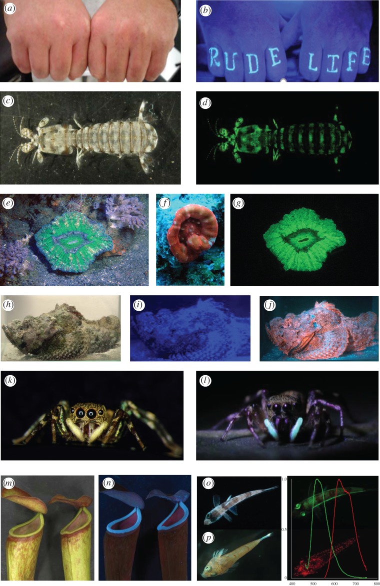
Fluorescence is a physico-chemical energy exchange where shorter-wavelength photons are absorbed by a molecule and are re-emitted as longer-wavelength photons. It has been suggested a means of communication in several taxa including flowers, pitcher plants, corals, algae, worms, squid, spiders, stomatopods, fish, reptiles, parrots and humans. The surface or object that the pigment molecule is part of appears to glow due to its setting rather than an actual production of light, and this may enhance both signals and, in some cases, camouflage. This review examines some known uses of fluorescence, mainly in the context of visual communication in animals, the challenge being to distinguish when fluorescence is a functional feature of biological coloration or when it is a by-product of a pigment or other molecule. In general, we conclude that most observations of fluorescence lack enough evidence to suggest they are used in visually driven behaviours.This article is part of the themed issue 'Animal coloration: production, perception, function and application'.

Keywords: behaviour; colour; excitation and emission; fluorescence; light; visual ecology.


Conflict of interest statement

We declare we have no competing interests.

Figures

Figure 1.
Figure 1.
Examples of fluorescent objects. (a,b) Fist tattoo that only ‘pops out’ under UV ‘black-light’ illumination, such as that used in nightclubs (http://itattoos.club/index.htm). (c,d) Stomatopod crustacean Lysiosquilla maculata under white light and blue (470 nm) excitation lamp. (e,f,g) Scolymia sp. solitary corals, (e,g) in situ under white and 470 nm peak flash illumination + 500 nm long-pass barrier filter, (f) at 17 m in ambient illumination with camera white balance on automatic. This photograph shows orange fluorescence and appears approximately as it does to unfiltered or light-boosted human eye. (h,i,j) Scorpaenid under white, simulated blue light of depth using 470 nm illumination and blue + 500 nm long-pass barrier filter (see figure 2i). Note the weak fluorescence of the fish cannot be seen when ‘washed out’ by blue alone. (k,l) Female jumping spider C. umbratica in white light and under UV excitation showing fluorescing pedipalps (photographs, D. Li). (m,n) Pitcher plant traps of Nepenthes in white light and under UV (366 nm peak) excitation (photographs, Anil J. Johnson, R. Kurup and S. Baby). (o) Green fluorescent deep-sea fish Chlorophthalmus. (p) Red fluorescing dragonet fish, graph shows normalized emission spectra per nm for each fish colour matched (photographs, M. Matz).
Figure 2.
Figure 2.
Fluorescence characteristics of the budgerigar M. undulatus (a–d) and the stomatopod crustacean L. glabriuscula (e–i). (a,b) White light and UV fluorescent excitation photographs of front and back of head showing fluorescent cheek and crown feathers. Excitation source was a ‘black light’. Shaded box indicates spectral zone removed by sunblock to remove fluorescence. (c) Normalized reflectance—lines colour-coded to approximately match bird; fluorescent crown feathers—light yellow, lower cheek, non-fluorescent yellow feathers—dark yellow (note difference in UV absorption of fluorescent feathers, which absorb in this wavelength range to re-emit as fluorescence), blue cheek patches—blue (note high UV reflectivity of these feathers and high chromatic contrast to yellow fluorescent feathers). UV ‘black-light’ excitation source used in (b)—solid black line, fluorescent excitation spectrum—long-dashed line, fluorescent emission—short-dashed line. (d) Spectral sensitivities—solid black lines, spectral emission difference (relative photons) between fluorescent feathers with and without sunblock applied—yellow line. (e,f) Frontal aggressive display in white light and blue-light excited fluorescence. (g) L. glabriuscula looking out from burrow in sand under fluorescent excitation. Fluorescent areas on antennal scales are conspicuous. (h) Fluorescent excitation spectrum—long-dashed line, fluorescent emission—short-dashed line. Spectral sensitivity of the row 2 distal photoreceptor in stomatopod retina showing excellent match to fluorescent emission range. (i) Ambient light in ocean at around 10 m in stomatopod habitat—blue line, fluorescent excitation lamp relative output—purple line and yellow blocking filter—yellow line. These were used in photography of stomatopods and match both natural ambient light and long–waveband-pass retinal filter of stomatopod (cryosection—natural colour—of filter inset photograph).

References

    1. Lagorio MG, Cordon GB, Iriel A. 2015. Reviewing the relevance of fluorescence in biological systems. Photochem. Photobiol. Sci. 14, 1538–1559. (10.1039/C5PP00122F) - DOI - PubMed
    1. Salih A, Larkum A, Cox G, Kuhl M, Hoegh-Guldberg O. 2000. Fluorescent pigments in corals are photoprotective. Nature 408, 850–853. (10.1038/35048564) - DOI - PubMed
    1. Johnsen S. 2012. The optics of life: a biologist's guide to light in nature. Princeton, NJ: Princeton University Press.
    1. Benson RC, Vogel MJ. 1955. The principles of identification and measurement of vulvar fluorescence. J. Clin. Endocrinol. Metab. 15, 784–800. (10.1210/jcem-15-7-784) - DOI - PubMed
    1. Weagle G, Paterson PE, Kennedy J, Pottier R. 1988. The nature of the chromophore responsible for naturally-occurring fluorescence in mouse skin. J. Photochem. Photobiol. B 2, 313–320. (10.1016/1011-1344(88)85051-6) - DOI - PubMed

LinkOut - more resources