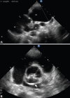
Bicuspid Aortic Valve: An Unusual Cause of Aneurysm of Left Coronary Sinus of Valsalva
- PMID: 28533581
- PMCID: PMC5429501
Bicuspid Aortic Valve: An Unusual Cause of Aneurysm of Left Coronary Sinus of Valsalva
Abstract
Bicuspid aortic valve is traditionally considered an innocuous congenital anomaly. Due to a better and widespread availability of non-invasive imaging techniques, it has come to the fore that 30% of these cases develop complications, viz., valve abnormality (aortic regurgitation and stenosis), and aneurysm of aortic root and ascending aorta. Sinus of Valsalva aneurysm is an uncommon complication of bicuspid aortic valve and more so those arising from the left coronary sinus are the rarest. These complications generally occur in the third or fourth decade of life. We present a case of the left sinus of Valsalva aneurysm in conjunction with bicuspid aortic valve and ascending aorta aneurysm at a very young age in a girl in her early adolescence. This case is to remind the paediatricians about the not so "innocuous image", but the serious implications of the bicuspid aortic valve and to regularly follow these cases for early diagnosis of potential complications so as to prevent catastrophic outcomes.
Keywords: Aortic aneurysm; Bicuspid aortic valve; Sinus of valsalva.
Figures


References
-
- Ward C. Clinical significance of the bicuspid aortic valve. Heart. 2000;83:81–5. [ PMC Free Article] - PMC - PubMed
-
- Kieffer SA, Winchell P. Congenital aneurysms of the aortic sinuses with cardioaortic fistula. Dis Chest. 1960;38:79–96. - PubMed
Publication types
LinkOut - more resources
Full Text Sources
