

Case Reports
doi: 10.4103/0972-3919.202233.
Unusual Presentation of Rare Cardiac Tumor: The Role of F-18-Fluorodeoxyglucose Positron Emission Tomography/Computed Tomography
Affiliations
- PMID: 28533653
- PMCID: PMC5439190
- DOI: 10.4103/0972-3919.202233
Item in Clipboard
Case Reports
Unusual Presentation of Rare Cardiac Tumor: The Role of F-18-Fluorodeoxyglucose Positron Emission Tomography/Computed Tomography
Indian J Nucl Med.
2017 Apr-Jun.
Abstract
Primary cardiac tumors are rare with angiosarcoma being the most common among malignant cardiac tumor. We present a case of 30-year-old female patient in whom F-18-fluorodeoxyglucose positron emission tomography/computed tomography demonstrated a necrotic mass in right atrium with multiple fluorodeoxyglucose avid lesions in both upper and lower alveolus, liver, multiple bones, and bilateral lungs. Patient underwent biopsy from gum swelling which revealed metastatic angiosarcoma.
Keywords: Angiosarcoma; F-18-fluorodeoxyglucose positron emission tomography/computed tomography; gingival metastasis; primary cardiac tumor.
Conflict of interest statement
There are no conflicts of interest.
Figures
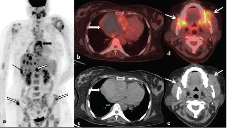
Whole body F-18 FDG PET/CT: (a) Maximum intensity projection demonstrated FDG uptake in the region of heart (thick arrow) and multiple foci of increased tracer uptake involving liver (thin arrow) and skeletal sites (outlined arrow). (b,c) Transaxial fused and CT images at the level of thorax showed large necrotic mass lesion in the region of right atrium with increased radiotracer uptake in the periphery (thick arrow). (d,e) Head and neck transaxial fused and CT images showed irregular soft tissue density lesion with increased FDG uptake involving bilateral alveolar region with subtle necrosis.
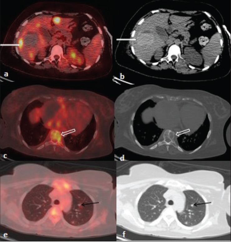
F-18 FDG PET/CT transaxial fused and CT images: (a, b) Transaxial images at the level of liver showed multiple foci of increased tracer uptake and large hyperdense mass lesion in segment V/VI of right lobe with mildly increased tracer uptake in the periphery (thin white arrow). (c-f) Transaxial images showed lytic lesions in multiple vertebrae (thick arrow) with increased radiotracer uptake and multiple nodules in bilateral lungs (thin black arrow) suggestive of metastases.
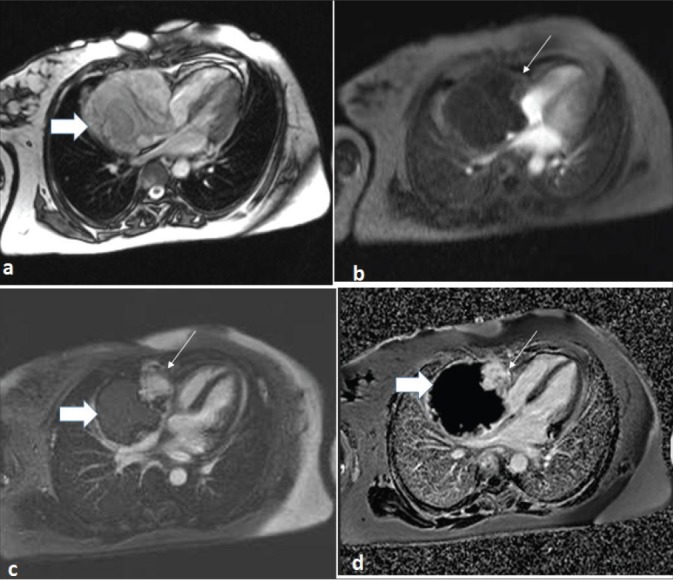
Cardiac magnetic resonance images: (a) Large lobulated intracavitary right atrial (RA) mass lesion with RA free wall appearing heterogenous on T2WI- 4CH TRUFI (four chamber true fast imaging with steady-state free precession). (b) perfusion images showed nodular uptake of contrast predominantly in the periphery (thin arrow) with no significant enhancement in rest of the mass (thick arrow). (c,d) Post GAD/PSIR images at 5 min demonstrated patchy nodular intense enhancement (thin arrow).
References
-
- Burke A, Virmani S. Atlas of tumor pathology. Washington, DC: Washington Armed Force Institute of Tumor Pathology; 1996.
-
- Reynen K. Cardiac myxomas. N Engl J Med. 1995;333:1610–17. - PubMed
-
- Kambiz R, Harald S, Michael S, Lars S, Andreas H, Tilmann S, et al. Differentiation of malignant and benign cardiac tumors using 18F-FDG PET/CT. J Nucl Med. 2012;53:856–63. - PubMed
-
- Bilski M, Kaminski G, Dziuk M. Metabolic activity assessment of cardiac angiosarcoma by 18FDG PET-CT. Nucl Med Rev Cent East Eur. 2012;15:83–4. - PubMed
Publication types
LinkOut - more resources
Full Text Sources
Other Literature Sources
