Review
. 2017 May 8:11:78.
doi: 10.3389/fncel.2017.00078. eCollection 2017.

Neonatal Hypoxia Ischaemia: Mechanisms, Models, and Therapeutic Challenges

Affiliations
Review

Neonatal Hypoxia Ischaemia: Mechanisms, Models, and Therapeutic Challenges

Lancelot J Millar et al. Front Cell Neurosci. .

Abstract

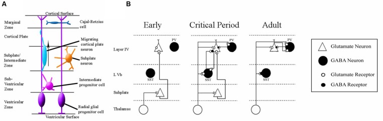
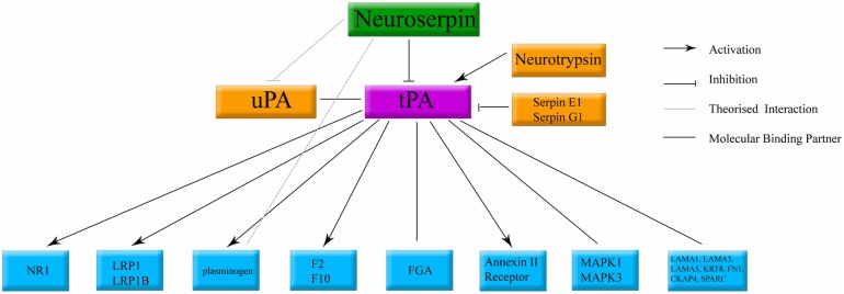
Neonatal hypoxia-ischaemia (HI) is the most common cause of death and disability in human neonates, and is often associated with persistent motor, sensory, and cognitive impairment. Improved intensive care technology has increased survival without preventing neurological disorder, increasing morbidity throughout the adult population. Early preventative or neuroprotective interventions have the potential to rescue brain development in neonates, yet only one therapeutic intervention is currently licensed for use in developed countries. Recent investigations of the transient cortical layer known as subplate, especially regarding subplate's secretory role, opens up a novel set of potential molecular modulators of neonatal HI injury. This review examines the biological mechanisms of human neonatal HI, discusses evidence for the relevance of subplate-secreted molecules to this condition, and evaluates available animal models. Neuroserpin, a neuronally released neuroprotective factor, is discussed as a case study for developing new potential pharmacological interventions for use post-ischaemic injury.

Keywords: encephalopathy; hypoxia-ischemia; neonatal; neurodevelopment; neuroprotection; neuroserpin; subplate.


Figures

FIGURE 1
FIGURE 1
Summary of Clinical Impact of Neonatal HI. This figures summarizes the number of neonates affected by neonatal HI annually across the globe relative to the number of live births per annum. Estimated figures for persistent disability associated with neonatal HI are also included. Estimates are based on data from studies references in this review (Hack et al., 1992; Vohr et al., 2000; Dilenge et al., 2001; Volpe, 2001, 2012; Graham et al., 2008; Lee et al., 2013).
FIGURE 2
FIGURE 2
Simplified Schematic of Brain Damage in Neonatal HI, approximately at the level of primary somatosensory and motor cortex. The two main patterns of injury, partially overlapping in patients, are shown separately for this schematic (adapted from Budday et al., 2014). Two colours have been used to show that many neonatal HI injuries consist of a centre of necrosis (black) and a penumbra of less acutely damaged tissue (gray). The exact location of these sites will vary depending on the nature of the injury. Black/gray areas represent the potential site of lesions, although those shown in this schematic are severe yet unilateral. (A) Primary basal-ganglia and thalamus injury pattern. (B) Primary watershed cortex and underlying white matter injury. Injury can primarily occur either to cortical gray matter or subcortical white matter depending on the nature of the injury. Severity also varies substantially between patients and within the brain of individuals. These have been documented in many human structural imaging studies (Barkovich et al., 1995; Cowan et al., 2003; Kaufman et al., 2003; Rutherford et al., 2004; Miller et al., 2005; Chau et al., 2008, 2012, 2013; Li et al., 2009).
FIGURE 3
FIGURE 3
Simplified Schematic of Major Molecular Injury Cascades involved in Neonatal HI. A vast number of molecules contribute to neonatal HI injury in the brain and all of these have been investigated in this disorder. These molecular targets can largely be split into the following three cascades. (A) Excitotoxicity (adapted from Vandenberg and Ryan, 2013) Ca2+ = calcium ion, Mg2+ = magnesium ion AMPA-R = α-amino-3-hydroxy-5-methyl-4-isoxazole propionic acid receptor, NMDA-R = N-methyl-D-aspartate receptor. (B) Oxidative Stress (adapted from http://www.enzolifesciences.com/platforms/cellular-analysis/oxidative-stress/). For simplicity, only free radicals are shown and physiological pathway substrates have been omitted. NADPH + Nicotinamide Adenine Dinucleotide Phosphate Hydrogen, O2 = oxygen, O2- = negatively-charged oxygen free radiccal, H2O2 = hydrogen peroxide, ROS = ROS1 receptor tyrosine kinase encoded by the ROS1 gene, Blc = B cell leukaemia protein, ER = endoplasmic reticulum, DNA = deoxyribose nucleic acid. (C) Inflammation (adapted from Santiago et al., 2014). BBB = blood brain barrier, IL-6 = interleukin 6, IL-1B = interleukin 1 beta, TNFalpha = tumour necrosis factor alpha, NO = nitric oxide.
FIGURE 4
FIGURE 4
Currently Used Rodent Models of Neonatal HI. (A) The Rice–Vannucci method as outlined in rats by Rice et al. (1981). (B) The general method with specifications taken from the pool of papers summarized in Supplementary Table 1. (C) The range of inflammatory models reviewed in Dean et al. (2015).
FIGURE 5
FIGURE 5
Schematics of the anatomy and physiology of human subplate development. (A) Schematic coronal section showing the major cellular compartments within the developing human cortex at 26 post-conception weeks, reviewed in Hoerder-Suabedissen and Molnár (2015). The germinal zone (site of cell division) consists of the ventricular zone and subventricular zone. The subplate and intermediate zone lie between the germinal zone and the cortical plate (the site into which the permanent layers will migrate). The outermost layer is the early-born marginal zone. (B) Classical schematic of subplate architecture throughout development. In the earliest phase of thalamocortical circuit establishment subplate neurons (white) receive inputs from thalamus, and project axons to layer 4. At the onset of critical period, both subplate neurons and thalamus project to layer 4. In adult, subplate neurons have been eliminated by programmed cell death and layer 4 neurons receive inputs directly from thalamus. Adapted from Kanold (2009). This classical view has been augmented by the inclusion of GABA-ergic layer Vb interneurons which have been demonstrated to contribute to thalamocortical circuitry development in somatosensory mouse cortex (Marques-Smith et al., 2016).
FIGURE 6
FIGURE 6
Classical binding cascade for neuroserpin protein. The main molecular target of neuroserpin is inhibition of tPA. Confirmed molecular binding partners are shown with a black line. Arrows denote confirmed activation. Orthogonal lines denote confirmed inactivation. Lines without caps denote confirmed binding with no documented physiological data. tPA, tissue plasminogen activator; uPA, urokinase plasminogen activator. All other abbreviations denote standard gene names.

References

    1. Adamsons K., Myers R. E. (1973). Perinatal asphyxia, causes, detection and neurologic sequelae. Pediatr. Clin. North Am. 20 465–480. 10.1016/S0031-3955(16)32855-3 - DOI - PubMed
    1. Ådén U., Halldner L., Lagercrantz H., Dalmau I., Ledent C., Fredholm B. B. (2003). Aggravated brain damage after hypoxic ischemia in immature adenosine A2A knockout mice. Stroke 34 739–744. 10.1161/01.STR.0000060204.67672.8B - DOI - PubMed
    1. Adhami F., Yu D., Yin W., Schloemer A., Burns K. A., Liao G., et al. (2008). Deleterious effects of plasminogen activators in neonatal cerebral hypoxia-ischemia. Am. J. Pathol. 172 1704–1716. 10.2353/ajpath.2008.070979 - DOI - PMC - PubMed
    1. Agrawal V., Hirsch E. (2012). Intrauterine infection and preterm labor. Semin. Fetal Neonatal Med. 17 12–19. 10.1016/j.siny.2011.09.001 - DOI - PMC - PubMed
    1. al Naqeeb N., Edwards A. D., Cowan F. M., Azzopardi D. (1999). Assessment of neonatal encephalopathy by amplitude-integrated electroencephalography. Pediatrics 103 1263–1271. 10.1542/peds.103.6.1263 - DOI - PubMed

LinkOut - more resources