. 2017 May 19:14:26.
doi: 10.1186/s12983-017-0212-2. eCollection 2017.

TC003132 is essential for the follicle stem cell lineage in telotrophic Tribolium oogenesis

Affiliations

TC003132 is essential for the follicle stem cell lineage in telotrophic Tribolium oogenesis

Matthias Teuscher et al. Front Zool. .

Abstract

Background: Stem cells are undifferentiated cells with a potential for self-renewal, which are essential to support normal development and homeostasis. To gain insight into the molecular mechanisms underlying adult stem cell biology and organ evolution, we use the telotrophic ovary of the beetle Tribolium. To this end, we participated in a large-scale RNAi screen in the red flour beetle Tribolium, which identified functions in embryonic and postembryonic development for more than half of the Tribolium genes.

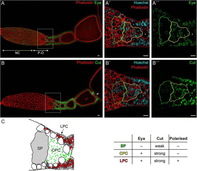
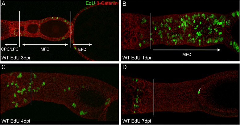
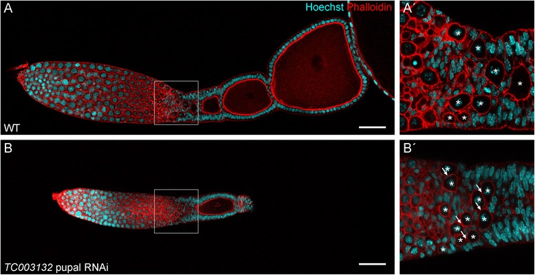
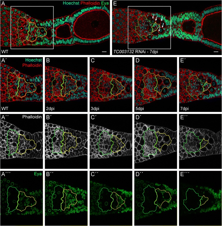
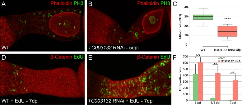
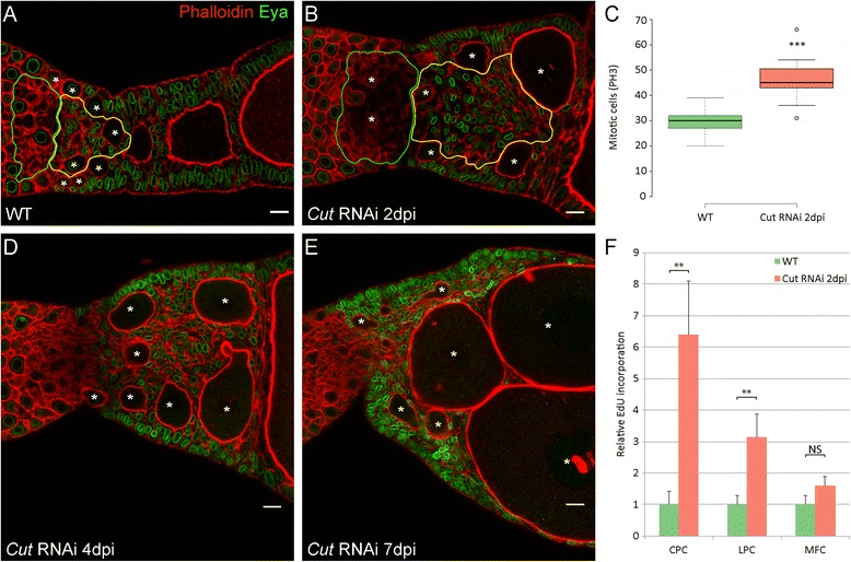
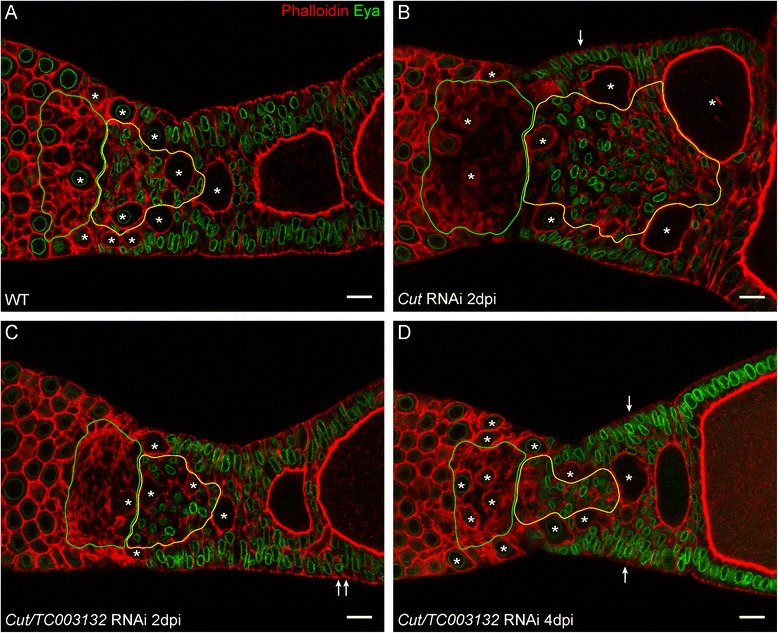
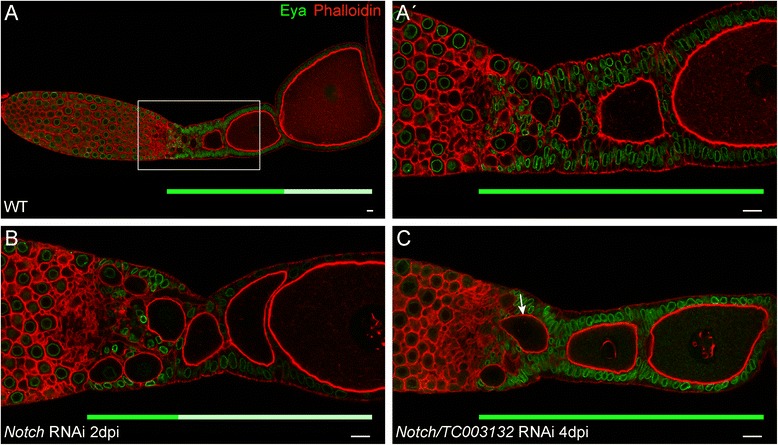
Results: We identified TC003132 as candidate gene for the follicle stem cell linage in telotrophic Tribolium oogenesis. TC003132 belongs to the Casein Kinase 2 substrate family (CK2S), which in humans is associated with the proliferative activity of different cancers. Upon TC003132 RNAi, central pre-follicular cells are lost, which results in termination of oogenesis. Given that also Notch-signalling is required to promote the mitotic activity of central pre-follicular cells, we performed epistasis experiments with Notch and cut. In addition, we identified a putative follicle stem cell population by monitoring the mitotic pattern of wild type and TC003132 depleted follicle cells by EdU incorporations. In TC003132 RNAi these putative FSCs cease the expression of differentiation makers and are eventually lost.

Conclusions: TC003132 depleted pre-follicular cells neither react to mitosis or endocycle stimulating signals, suggesting that TC003132 provides competence for differentiation cues. This may resemble the situation in C. elegans were CK2 is required to maintain the balance between proliferation and differentiation in the germ line. Since the earliest effect of TC003132 RNAi is characterized by the loss of putative FSCs, we posit that TC003132 crucially contributes to the proliferation or maintenance of follicle stem cells in the telotrophic Tribolium ovary.

Keywords: Notch-signalling; Tribolium; follicle stem cells; iBeetle; large-scale RNAi screen; telotrophic oogenesis.


Figures

Fig. 1
Fig. 1
(AB″) Detection of Eya (AA″) and Cut (BB″) (green) Phalloidin for f-Actin (red) and Hoechst 33,258 for DNA (blue) in wild type ovarioles. A′A″ and B′B″ are magnifications of boxed regions in A and B, respectively. The asterisk in AA″ marks the first aligned oocyte. (C) Schematic representation and features of somatic cell types of boxed regions in A and B. See text for details. NC: nurse cells; P-O: pro-oocytes; SP: somatic plug; CPC: central prefollicular cells; LPC: lateral prefollicular cells; green and yellow outlines mark SP and CPCs, respectively. Scale Bar: 10 μm
Fig. 2
Fig. 2
ad Detection of EdU (green) and ß-Catenin (red) in wild type ovarioles. In (a) a single focal plane is shown two visualize the distinction between the different cell populations. bd are standard deviation projections reflecting total EdU incorporation. The borders between CPCs/LPCs and MFCs are indicated by a solid white bar. See text for further details. CPC: central prefollicular cells; LPC: lateral prefollicular cells; MFC: mitotic follicle cells; EFC: endocycling follicle cells; dpi: days post injection
Fig. 3
Fig. 3
(AA′) Wild type ovariole of Tribolium stained with Phalloidin for f-Actin (red) and Hoechst (blue). In wild type, pro-oocytes leave the tropharium, become encapsulated by follicle cells and are aligned in the centre (A′, magnification of indicated region in A). (BB′) Pupal knockdown of TC003132 at 7dpi. The vitellarium is shortened and only a single growing follicle is obvious (B). Oocytes are not longer encapsulated, resulting in oocytes arranged side-by-side (B′). Asterisks mark oocytes and Arrows point to oocytes contacting each other, thus indicating encapsulation defects. Scale bar in A and B: 50 μm
Fig. 4
Fig. 4
(AA‴) Wild type ovariole stained with Phalloidin for f-Actin (red in AA′, white in A″), Eya (green), and Hoechst (blue). Eya is expressed in central pre-follicular cells (CPC, yellow outline) and the somatic plug (SP, green outline) (BE‴) Ovarioles dissected at indicated time points after adult knockdown of TC003132. A subset of CPC starts to express Eya at lower levels (border between high and low expression of Eya in CPCs is marked by the yellow dashed line) (BB″). At 3dpi, CPCs occupy a smaller area and Eya expression is further reduced. The somatic plug (SP) appears less condensed and occupies a larger area (CC″). CPCs expressing reduced amounts of Eya are lost at 5dpi (DD″). The terminal phenotype can be observed at 7dpi. Only a small population of Eya positive CPCs is still present and the SP has expanded even more. Also encapsulation defects become apparent as young oocytes can be found in direct contact to each other (arrows, EE‴). Scale bar: 10 μm
Fig. 5
Fig. 5
ab Staining of phosphorylated histon H3 (PH3, green) and Phalloidin (red) in wild type and TC003132 RNAi ovarioles. c Box plot of PH3 positive cells in WT (green) and TC003132 RNAi at 5dpi (red). Centre lines show the medians; box limits indicate the 25th and 75th percentiles as determined by R software; whiskers extend 1.5 times the interquartile range from the 25th and 75th percentiles. n = 13, 14 sample points. df Detection of EdU (green) and ß-Catenin (red) in wild type and TC003132 RNAi ovarioles. Compared to wild type, the amount of follicle cells in mitosis is approximately halved 5dpi after TC003132 RNAi (ac). Both, wild type and TC003132 knockdown exhibit similar amounts of EdU incorporation at 3dpi (f). In wild type, the signal is rapidly lost in (d, f) and only the anteriormost central pre-follicular cells remain EdU positive (d). In TC003132 RNAi, the EdU signal is stable and diminishes at a much slower rate (e, f). Error bars in (f) represent standard deviation. Dpi: days post injection; NS: Not significant; ** and **** P < 0.01 and P < 0.0001 using a two-tailed t-test, respectively
Fig. 6
Fig. 6
a Wild type ovariole stained with Phalloidin to visualize f-Actin (red) and an antibody against Eya (green). b, d, e RNAi phenotypes of Cut at indicated time points. The Knockdown of Cut results in the over proliferation of follicle cell populations with CPCs showing the strongest effect (b, yellow outline). Oocytes are not longer aligned, but positioned of side-by-side (asterisks). At subsequent dissections continuous growth of oocytes can be observed, resulting in the dislocation of CPCs (de). c Box plot of PH3 positive cells in WT (green) and Cut RNAi at 2dpi (red) shows increased mitosis rate after Cut knockdown. Centre lines show the medians; box limits indicate the 25th and 75th percentiles as determined by R software; whiskers extend 1.5 times the interquartile range from the 25th and 75th percentiles, outliers are represented by dots. n = 13, 11 sample points. f EdU signals in WT and Cut RNAi normalized to the mean of wild type. Error bars represent standard deviation. Central pre-follicular cells show the highest relative EdU incorporation (f). ** and *** P < 0.01 and P < 0.001 using a two-tailed t-test, respectively. Scale bar: 10 μm
Fig. 7
Fig. 7
a Wildtype, b Cut RNAi, and cd Double knockdown of Cut and TC003132 at indicated time points. Two days after injection, only a mild cut phenotype is obvious. Minor over-proliferation can be observed (arrows, c). At 4dpi, multi-layered follicle cells (arrows) become more frequent, but other effects of Cut RNAi are supressed (d). Oocytes are marked by asterisks; Scale bar: 10 μm
Fig. 8
Fig. 8
(AA′) Wild type ovariole stained with Phalloidin to visualize f-Actin (red) and an antibody against Eya (green). Mitotic active follicle cells strongly express Eya, while cells that entered the endocycle cease Eya expression. (A′) Magnification of the indicated region in (A). (B) Knockdown of Notch results in the premature entry of the follicle cells into the endocycle. (C) Double RNAi of Notch and TC003132 suppresses the effects of Notch RNAi in follicle cells: Eya expression is restored. Still, the germline shows signs of the Notch phenotype as prematurely growing oocytes are still obvious (C, arrow). Solid green and light green bars below ovarioles indicate regions of strong and weak Eya expression, respectively. Scale bar: 10 μm

Similar articles

Cited by

References

    1. Kirilly D, Xie T. The Drosophila ovary: an active stem cell community. Cell Res. 2007;17:15–25. doi: 10.1038/sj.cr.7310123. - DOI - PubMed
    1. Losick VP, Morris LX, Fox DT, Spradling A. Drosophila stem cell niches: a decade of discovery suggests a unified view of stem cell regulation. Dev Cell. 2011;21:159–171. doi: 10.1016/j.devcel.2011.06.018. - DOI - PMC - PubMed
    1. Sahai-Hernandez P, Nystul TG. A dynamic population of stromal cells contributes to the follicle stem cell niche in the Drosophila ovary. Development. 2013;140:4490–4498. doi: 10.1242/dev.098558. - DOI - PMC - PubMed
    1. Margolis J, Spradling A. Identification and behavior of epithelial stem cells in the Drosophila ovary. Development. 1995;121:3797–3807. - PubMed
    1. Song X, Xie T. DE-cadherin-mediated cell adhesion is essential for maintaining somatic stem cells in the Drosophila ovary. Proc Natl Acad Sci. 2002;99:14813–14818. doi: 10.1073/pnas.232389399. - DOI - PMC - PubMed

LinkOut - more resources