Review
. 2017 Sep;134(3):331-349.
doi: 10.1007/s00401-017-1718-6. Epub 2017 May 22.

White matter injury in the preterm infant: pathology and mechanisms

Affiliations
Review

White matter injury in the preterm infant: pathology and mechanisms

Stephen A Back. Acta Neuropathol. 2017 Sep.

Abstract

The human preterm brain is particularly susceptible to cerebral white matter injury (WMI) that disrupts the normal progression of developmental myelination. Advances in the care of preterm infants have resulted in a sustained reduction in the severity of WMI that has shifted from more severe focal necrotic lesions to milder diffuse WMI. Nevertheless, WMI remains a global health problem and the most common cause of chronic neurological morbidity from cerebral palsy and diverse neurobehavioral disabilities. Diffuse WMI involves maturation-dependent vulnerability of the oligodendrocyte (OL) lineage with selective degeneration of late oligodendrocyte progenitors (preOLs) triggered by oxidative stress and other insults. The magnitude and distribution of diffuse WMI are related to both the timing of appearance and regional distribution of susceptible preOLs. Diffuse WMI disrupts the normal progression of OL lineage maturation and myelination through aberrant mechanisms of regeneration and repair. PreOL degeneration is accompanied by early robust proliferation of OL progenitors that regenerate and augment the preOL pool available to generate myelinating OLs. However, newly generated preOLs fail to differentiate and initiate myelination along their normal developmental trajectory despite the presence of numerous intact-appearing axons. Disrupted preOL maturation is accompanied by diffuse gliosis and disturbances in the composition of the extracellular matrix and is mediated in part by inhibitory factors derived from reactive astrocytes. Signaling pathways implicated in disrupted myelination include those mediated by Notch, WNT-beta catenin, and hyaluronan. Hence, there exists a potentially broad but still poorly defined developmental window for interventions to promote white matter repair and myelination and potentially reverses the widespread disturbances in cerebral gray matter growth that accompanies WMI.

Keywords: Astrocyte; Development; Dysmaturation; Glia; Hypoxia–ischemia; MRI; Microglia; Oligodendrocyte; Periventricular leukomalacia; Prematurity.


Figures

Figure 1
Figure 1. Severe WMI results in focal macroscopic or microscopic necrosis
(A) Gross pathology demonstrates the typical features of severe cystic necrotic WMI in an infant who died of complications of prematurity. Note the large foci of severe cystic necrosis (arrowheads) in frontal (upper specimen) and parietal (lower specimens) periventricular white matter. (B) Histological analysis of the frontal lesion (stained with hematoxylin and eosin) shows a large focus of necrosis (arrowheads) adjacent to the external angle of lateral ventricle. The inset shows a high power detail of the edge of the lesion (arrows) where marked rarefaction of the tissue can be appreciated adjacent to a region of gliosis at lower left. (C) Appearance of cystic necrotic WMI on MRI. Images from a preterm infant born at 33 weeks gestational age and scanned at 5 weeks of age (38 weeks adjusted gestational age). Small areas of cavitation (arrowheads) are appreciated as hypointensity on the sagittal T1 weighted image, and as hyperintensity on the axial T2 weighted image, (D, E) Low power image of the typical sparse and focal distribution of microscopic necrosis in the periventricular white matter from a human autopsy brain at 32 weeks post conceptional age. The microcyst was visualized with β-amyloid precursor protein, a marker of degenerating axons. (F, G) Panels F and G provide higher power detail of the degenerating axons in the microcyst seen in the box in E and demonstrate that axonal degeneration was present both within the core and periphery of the microcyst. Scale bars: E, 500 μm; F, 100 μm; G, 25 μm. (Images in A and B, Courtesy of Dr. Marjorie Grafe, Oregon Health & Science University. C, D, Courtesy of Dr. Ken Poskitt, Children’s and Women’s Hospital, University of British Columbia; E-F adapted from Back and Miller, 2014; [19]).
Figure 2
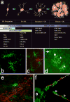
Figure 2. The differentiation of human oligodendrocyte (OL) progenitors and myelination progresses according to a well-defined lineage defined by specific markers
(A) Diagram depicting the maturation of the OL lineage. The four principal stages of the OL lineage are depicted together with their corresponding morphological features and potential for proliferation, migration or myelination. Each stage is uniquely defined by a combination of marker genes or antibodies. A2B5, O4, O1 refer to mouse monoclonal antibodies. Olig 2 and Sox10 are genes that are highly enriched in premyelinating OL progenitors. Olig 2 is also expressed at later stages in the OL lineage. Abbreviations: CNP (CNPase), 2′:3′-cyclic nucleotide-3′-phosphodiesterase; GalC, galactocerebroside; MAG, myelin associated glycoprotein; MBP, myelin basic protein; MOG, myelin oligodendrocyte glycoprotein; NG2, chondroitin sulfate proteoglycan 4; PDGFRα, platelet-derived growth factor-alpha; PLP, proteolipid protein. (B, C) Human preOLs (green) visualized with the O4 antibody. In C, note the GFAP-labeled astrocyte (red) with distinctly different morphology from the preOLs (green). (D) Human immature OLs visualized with the O4 antibody (arrowheads), which appear much more ramified compared to adjacent preOLs (arrows). (E) An early myelinating human immature OL (green; O1 antibody-labeled) extends several fine processes that make apparent contacts with individual axons visualized with the pan-axonal neurofilament protein marker, SMI312. (F) A 3-dimensional reconstruction of an axon in the early stages of myelination that was visualized in the optic radiation from a case at 30-weeks post-conceptional age. An early myelinating OL, visualized with the O4 antibody, generates early loose axonal contacts (yellow) and myelin sheath wrappings (green) that spiral around an axon visualized with SMI312 (red). Two views of the axon are shown. The portion of the axon visualized at lower right demonstrates that the myelin sheath appears to be laid down in a segmental fashion (arrowheads). A portion of the O4-labeled OL soma is seen at lower left (s). The images in E and F were adapted from Back et. al., 2002 [17].
Figure 3
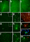
Figure 3. Acute human diffuse WMI is accompanied by selective preOL death
(A-C) Cell death is visualized in A-C by TUNEL staining, which detects DNA fragmentation of apoptotic and necrotic cells. A, Rare cell death was seen in the overlying cerebral cortex, shown here from analysis of a preterm infant with diffuse WMI at autopsy. The arrow marks the pial surface. (B, C) The periventricular white matter from the same case showed extensive cell death that was confirmed below to be due to preOL degeneration. The individual degenerating cells appear as bright green. (D-F) Typical example of a gradient of early WMI that preferentially localized to deeper areas of cerebral white matter in D and E with more normal appearing white matter in F. Note the marked reduction in staining for preOLs in D and E compared to F. Also note the severely damaged cell remnants in D (arrow; detail in inset) and the paucity of normal appearing preOLs in E (arrow; detail in inset). (G) Typical appearance of shrunken degenerating preOLs (arrows) scattered among more normal-appearing cells (arrowheads), also shown in the inset. (H, I) Typical morphology of a degenerating preOLs with a halo of fragmented degenerating processes. (J) Numeous preOLs (red) also seen adjacent to but not within a focal lesion (arrows) in the subventricular zone deep to the periventricular white matter. Note the many degenerating TUNEL-labeled cells (green). (K-M) Examples of apoptotic preOLs with a typical fragmented condensed nucleus. Adapted from reference: [18] Scale bars: A, 200 μm; B, 100 μm; C, 200 μm; D-F, 50 μm; Inset in D and E, 10 μm; G and inset in G, 50 μm; H, 10μm; I, 20 μm; J, 200 μm; K-M, 10μm.
Figure 4
Figure 4. Chronic WMI displays myelination disturbances related to arrested preOL maturation
(A) Distinctly different pathogenetic mechanisms mediate abnormal myelination in focal necrotic lesions (periventricular leukomalacia (PVL); upper pathway) versus lesions with diffuse WMI (lower pathway). When more severe, hypoxia–ischemia triggers white matter necrosis (upper pathway) with pancellular degeneration that depletes the white matter of glia and axons. Severe necrosis results in cystic PVL, whereas milder necrosis results in microcysts. Milder hypoxia–ischemia (lower pathway) selectively triggers early preOL death, but preOLs are rapidly regenerated in chronic lesions enriched in reactive astrocytes that contribute to a block in preOL differentiation to myelinating OLs. Myelination failure in diffuse WMI thus results from preOL arrest rather than axonal degeneration, as occurs with white matter necrosis. Note that the lower pathway is the dominant one for many contemporary preterm survivors, whereas the minor upper pathway reflects the declining burden of white matter necrosis. (B) Typical appearance of normal early myelination in neonatal rodents. Axons are visualized in red and early myelination of axons is in green. (C) Arrested maturation of preOLs in a chronic white matter lesion arising from hypoxia-ischemia (HI) where numerous preOLs (green; arrowheads) are seen, but the axons (red; arrows) are diffusely unmyelinated. (D) Normal early myelination (O1-antibody; green) in control subcortical white matter (corpus callosum/external capsule) at postnatal day10 (1 week after HI). There are low levels of GFAP-labeled astrocytes (red) in the white matter, adjacent cortex (CTX) and caudate putamen (CPu). (E) Absence of myelin (green) in the contralateral post-ischemic lesion coincided with a diffuse glial scar that stained diffusely for GFAP-labeled astrocytes (red). (F) Early myelination in control white matter at P10 with sheaths (yellow) double-labeled for O4 and O1 antibodies. (G) Absence of myelin in the contralateral lesion coincided with clusters of preOLs (O4+O1-) in maturation arrest (red; arrowheads). Such dense clusters of preOLs are not normally seen in control white matter and are consistent with the pronounced proliferative state that is triggered in response to injury. Apparent oligodendrocytes (yellow; arrows; O4+O1+) are rarely seen in the lesions. Panels A-C adapted from Back and Miller, 2014 [19]. Panels D-G adapted from Back and Rosenberg, 2014 [20]. Scale bars: B, C 100μm.

References

    1. (CDC) CfDCaP. Economic costs associated with mental retardation, cerebral palsy, hearing loss and vision impairment–United States, 2003. MMWR Morb Mortal Wkly Rep. 2004;53:57–59. - PubMed
    1. Aarnoudse-Moens CS, Weisglas-Kuperus N, van Goudoever JB, Oosterlaan J. Meta-analysis of neurobehavioral outcomes in very preterm and/or very low birth weight children. Pediatrics. 2009;124:717–728. - PubMed
    1. Aguirre A, Dupree JL, Mangin JM, Gallo V. A functional role for EGFR signaling in myelination and remyelination. Nat Neurosci. 2007;10:990–1002. - PubMed
    1. Alix JJ, Fern R. Glutamate receptor-mediated ischemic injury of premyelinated central axons. Ann Neurol. 2009;66:682–693. - PubMed
    1. Alix JJ, Zammit C, Riddle A, Meshul CK, Back SA, Valentino M, Fern R. Central axons preparing to myelinate are highly sensitivity to ischemic injury. Ann Neurol. 2012;72:936–951. - PMC - PubMed