

Microglia: Housekeeper of the Central Nervous System
- PMID: 28534246
- PMCID: PMC11481884
- DOI: 10.1007/s10571-017-0504-2
Microglia: Housekeeper of the Central Nervous System
Abstract
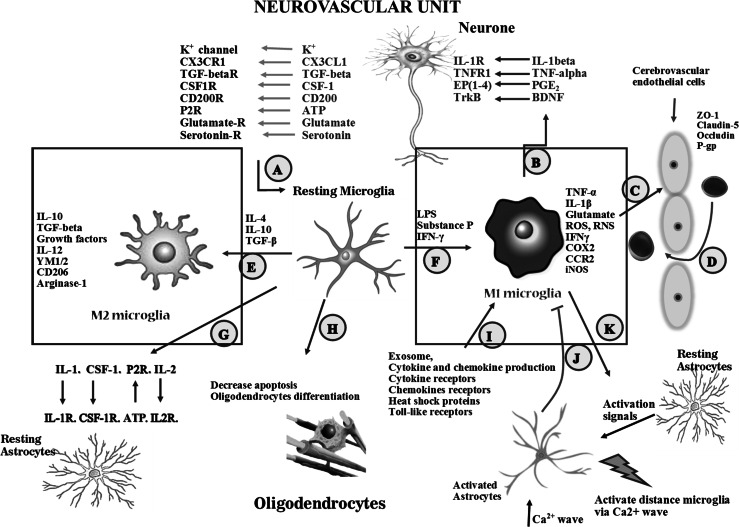
Microglia, of myeloid origin, play fundamental roles in the control of immune responses and the maintenance of central nervous system homeostasis. These cells, just like peripheral macrophages, may be activated into M1 pro-inflammatory or M2 anti-inflammatory phenotypes by appropriate stimuli. Microglia do not respond in isolation, but form part of complex networks of cells influencing each other. This review addresses the complex interaction of microglia with each cell type in the brain: neurons, astrocytes, cerebrovascular endothelial cells, and oligodendrocytes. We also highlight the participation of microglia in the maintenance of homeostasis in the brain, and their roles in the development and progression of age-related neurodegenerative disorders.
Keywords: Astrocytes; Brain aging; CNS keeper; Microglia activation; Neurons; Oligodendrocytes.
Conflict of interest statement
The authors declare that they have no conflict of interest for this manuscript.
Figures


Similar articles
-
Microglia and Macrophages in the Pathological Central and Peripheral Nervous Systems.Cells. 2020 Sep 21;9(9):2132. doi: 10.3390/cells9092132. Cells. 2020. PMID: 32967118 Free PMC article. Review.
-
Microglia and the Brain: Complementary Partners in Development and Disease.Annu Rev Cell Dev Biol. 2018 Oct 6;34:523-544. doi: 10.1146/annurev-cellbio-100616-060509. Epub 2018 Aug 8. Annu Rev Cell Dev Biol. 2018. PMID: 30089221 Review.
-
Microglia as Dynamic Cellular Mediators of Brain Function.Trends Mol Med. 2019 Nov;25(11):967-979. doi: 10.1016/j.molmed.2019.08.013. Epub 2019 Oct 6. Trends Mol Med. 2019. PMID: 31597593 Free PMC article. Review.
-
Cardiolipin in Central Nervous System Physiology and Pathology.Cell Mol Neurobiol. 2017 Oct;37(7):1161-1172. doi: 10.1007/s10571-016-0458-9. Epub 2016 Dec 30. Cell Mol Neurobiol. 2017. PMID: 28039536 Free PMC article. Review.
-
Microglia and Aging: The Role of the TREM2-DAP12 and CX3CL1-CX3CR1 Axes.Int J Mol Sci. 2018 Jan 22;19(1):318. doi: 10.3390/ijms19010318. Int J Mol Sci. 2018. PMID: 29361745 Free PMC article. Review.
Cited by
-
Extracellular Vesicles Cargo in Modulating Microglia Functional Responses.Biology (Basel). 2022 Sep 29;11(10):1426. doi: 10.3390/biology11101426. Biology (Basel). 2022. PMID: 36290330 Free PMC article.
-
Lutein Exerts Antioxidant and Anti-Inflammatory Effects and Influences Iron Utilization of BV-2 Microglia.Antioxidants (Basel). 2021 Feb 27;10(3):363. doi: 10.3390/antiox10030363. Antioxidants (Basel). 2021. PMID: 33673707 Free PMC article.
-
The association between cerebrospinal ferritin and soluble triggering receptor expressed on myeloid cells 2 along Alzheimer's continuum.Front Neurol. 2022 Nov 3;13:961842. doi: 10.3389/fneur.2022.961842. eCollection 2022. Front Neurol. 2022. PMID: 36408515 Free PMC article.
-
Synapse Dysfunctions in Multiple Sclerosis.Int J Mol Sci. 2023 Jan 13;24(2):1639. doi: 10.3390/ijms24021639. Int J Mol Sci. 2023. PMID: 36675155 Free PMC article. Review.
-
Sex- and Development-Dependent Responses of Rat Microglia to Pro- and Anti-inflammatory Stimulation.Front Cell Neurosci. 2018 Nov 20;12:433. doi: 10.3389/fncel.2018.00433. eCollection 2018. Front Cell Neurosci. 2018. PMID: 30524242 Free PMC article.
References
-
- Ahluwalia A, Jones MK, Szabo S, Tarnawski AS (2014) Aging impairs transcriptional regulation of vascular endothelial growth factor in human microvascular endothelial cells: implications for angiogenesis and cell survival. J Physiol Pharmacol 65:209–215 - PubMed
-
- Akundi RS, Candelario-Jalil E, Hess S, Hull M, Lieb K, Gebicke-Haerter PJ, Fiebich BL (2005) Signal transduction pathways regulating cyclooxygenase-2 in lipopolysaccharide-activated primary rat microglia. Glia 51:199–208. doi:10.1002/glia.20198 - PubMed
Publication types
MeSH terms
Grants and funding
LinkOut - more resources
Full Text Sources
Other Literature Sources
