. 2017 Jun 14;17(6):3932-3939.
doi: 10.1021/acs.nanolett.7b01523. Epub 2017 May 24.

Intracellular and Extracellular Recording of Spontaneous Action Potentials in Mammalian Neurons and Cardiac Cells with 3D Plasmonic Nanoelectrodes

Affiliations

Intracellular and Extracellular Recording of Spontaneous Action Potentials in Mammalian Neurons and Cardiac Cells with 3D Plasmonic Nanoelectrodes

Michele Dipalo et al. Nano Lett. .

Abstract

Three-dimensional vertical micro- and nanostructures can enhance the signal quality of multielectrode arrays and promise to become the prime methodology for the investigation of large networks of electrogenic cells. So far, access to the intracellular environment has been obtained via spontaneous poration, electroporation, or by surface functionalization of the micro/nanostructures; however, these methods still suffer from some limitations due to their intrinsic characteristics that limit their widespread use. Here, we demonstrate the ability to continuously record both extracellular and intracellular-like action potentials at each electrode site in spontaneously active mammalian neurons and HL-1 cardiac-derived cells via the combination of vertical nanoelectrodes with plasmonic optoporation. We demonstrate long-term and stable recordings with a very good signal-to-noise ratio. Additionally, plasmonic optoporation does not perturb the spontaneous electrical activity; it permits continuous recording even during the poration process and can regulate extracellular and intracellular contributions by means of partial cellular poration.

Keywords: Intracellular recording; cardiomyocytes; multielectrode arrays; neurons; plasmonic optoporation.


Conflict of interest statement

The authors declare no competing financial interest.

Figures
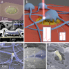
Figure 1
Figure 1
Schematic illustration of the plasmonic opto-operation platform integrated with a hippocampal neuronal culture-system. (a) Colored SEM image of an electrode with plasmonic 3D nanoelectrodes. (b) Representation of neurons on MEA with 3D nanoelectrodes. The 3D nanoelectrode excited with laser records intracellular activity, while the rest of the electrode catches extracellular signals. (c) Colored SEM cross-section of a neuronal process (blue) engulfing two 3D nanoelectrodes (yellow). The cross-section was obtained with focused ion beam milling using a top protecting platinum layer (identified with * in the image). The inset shows the top view of the neuronal process before ion milling. (d, e, f) Colored SEM images of neuronal soma and processes enveloping gold plasmonic 3D nanoelectrodes. The culture was fixed at 10 div.
Figure 2
Figure 2
Extracellular and intracellular-like firing activity recordings of hippocampal neurons before and after plasmonic optoporation. (a) Spontaneous extracellular activity of neurons at 20 DIV. (b) Intracellular-like spontaneous activity in the same neuron in panel a after optoporation. (c) Amplitude of the positive phase of action potentials after optoporation. (d) Spontaneous extracellular spike taken from the track in panel a where indicated with an asterisk. (e) Spontaneous intracellular-like spike recorded from the same neuron after optoporation. (f) Spontaneous intracellular-like and extracellular spikes recorded from two neurons on the same electrode, taken from panel b where indicated with an asterisk. (g) Experimental spike with 3D nanoelectrode inside the cell superimposed with SPICE simulation. (h, i) Pure intracellular (h) and extracellular (i) spikes extracted from the equivalent circuit obtained by fitting the experimental extra- and intracellular spikes (see the SI).
Figure 3
Figure 3
Characteristics of intracellular-like firing activity recordings in hippocampal neurons. (a) Primary neuron spontaneous activity with the presence of small positive peaks before the action potential. (b) Superimposition of small peaks that might be related to subthreshold activity. (c) Superimposition of spikes from the same neuron showing an initial bump. (d) Recording of intracellular-like spontaneous activity from two electrodes of the same MEA.
Figure 4
Figure 4
Extracellular and intracellular-like firing activity recordings of HL-1 cells before and after plasmonic optoporation. (a) Extracellular action potential of HL-1 cardiac cells cultured on MEA with 3D nanoelectrodes for 3 days. (b) Intracellular-like action potential of the same cell recorded in panel a after plasmonic optoporation was performed on the 3D nanoelectrodes underlying the cell. (c) Action potential of the same cell as in panel a and b more than 90 min after plasmonic optoporation; the cell membrane has reformed, and the spike shape closely resembles the initial extracellular spike before poration. (d) Recording of intracellular cardiac action potentials immediately after plasmonic optoporation performed on one 3D nanoelectrode. The amplitude of the intracellular spikes presents a gradual growth within the first 2 s after plasmonic poration. This phenomenon may reflect the real-time evolution of the membrane resettling on the nanoelectrode after the poration. (e) Recording of intracellular cardiac action potentials during further laser excitation of a nanoelectrode underlying an already porated cell. The red band indicates the time window of laser excitation (350 ms). An action potential is recorded while laser excitation is occurring without any alteration of the signal shape and the beating frequency immediately after laser excitation is unaltered.
Figure 5
Figure 5
Extracellular and intracellular contributions to the spikes of HL-1 cells after plasmonic optoporation. (a) Continuous recording of cardiac activity during the poration performed on four nanoelectrodes. The left part of the recording represents the extracellular activity. When two out of the four nanoelectrodes are used to porate the HL-1 cell, the signals acquire a hybrid shape that contains extra- and intracellular potentials (center region of the recording). When all four nanoelectrodes are intracellularly coupled, the signals acquire the more typical shape of the intracellular action potential. (b) Schematic view of an HL-1 cell lying on an electrode decorated by 4 3D nanoelectrodes. (c, d, e, f) Magnified view of cardiac action potentials according to how many 3D nanoelectrodes have been used for plasmonic poration; from left to right: 0, 1, 2, and 4 intracellular 3D nanoelectrodes. Red lines are actual recordings, and blue lines are simulations.

References

    1. Duan X.; Gao R.; Xie P.; Cohen-Karni T.; Qing Q.; Choe H. S.; Tian B.; Jiang X.; Lieber C. M. Nat. Nanotechnol. 2011, 7, 174–179. 10.1038/nnano.2011.223. - DOI - PMC - PubMed
    1. Xie C.; Lin Z.; Hanson L.; Cui Y.; Cui B. Nat. Nanotechnol. 2012, 7, 185–190. 10.1038/nnano.2012.8. - DOI - PMC - PubMed
    1. Lin Z. C.; Xie C.; Osakada Y.; Cui Y.; Cui B. Nat. Commun. 2014, 5, 3206.10.1038/ncomms4206. - DOI - PMC - PubMed
    1. Robinson J. T.; Jorgolli M.; Shalek A. K.; Yoon M.-H.; Gertner R. S.; Park H. Nat. Nanotechnol. 2012, 7, 180–184. 10.1038/nnano.2011.249. - DOI - PMC - PubMed
    1. Spira M. E.; Hai A. Nat. Nanotechnol. 2013, 8, 83–94. 10.1038/nnano.2012.265. - DOI - PubMed

Publication types