. 2017 May 23:8:15408.
doi: 10.1038/ncomms15408.

Rules of engagement between αvβ6 integrin and foot-and-mouth disease virus

Affiliations

Rules of engagement between αvβ6 integrin and foot-and-mouth disease virus

Abhay Kotecha et al. Nat Commun. .

Abstract

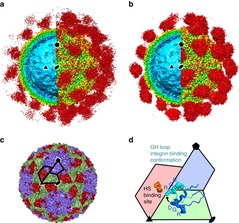
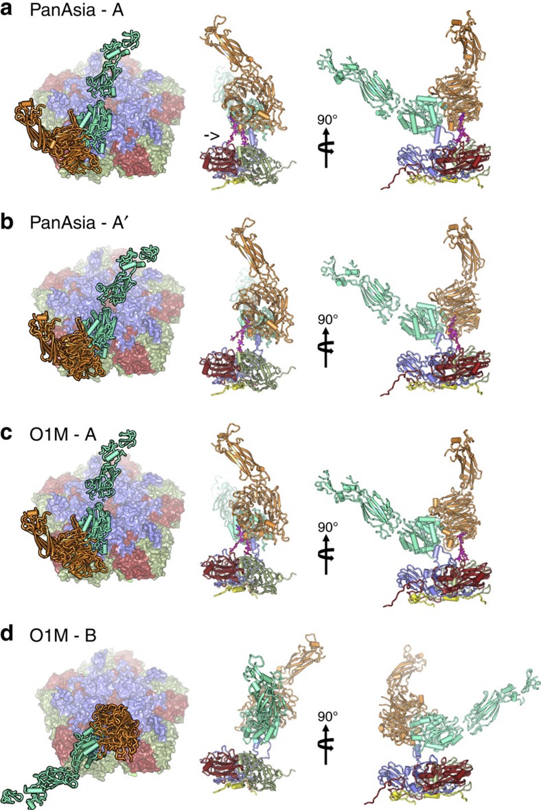
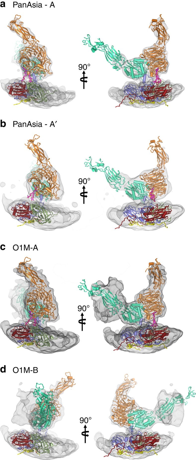
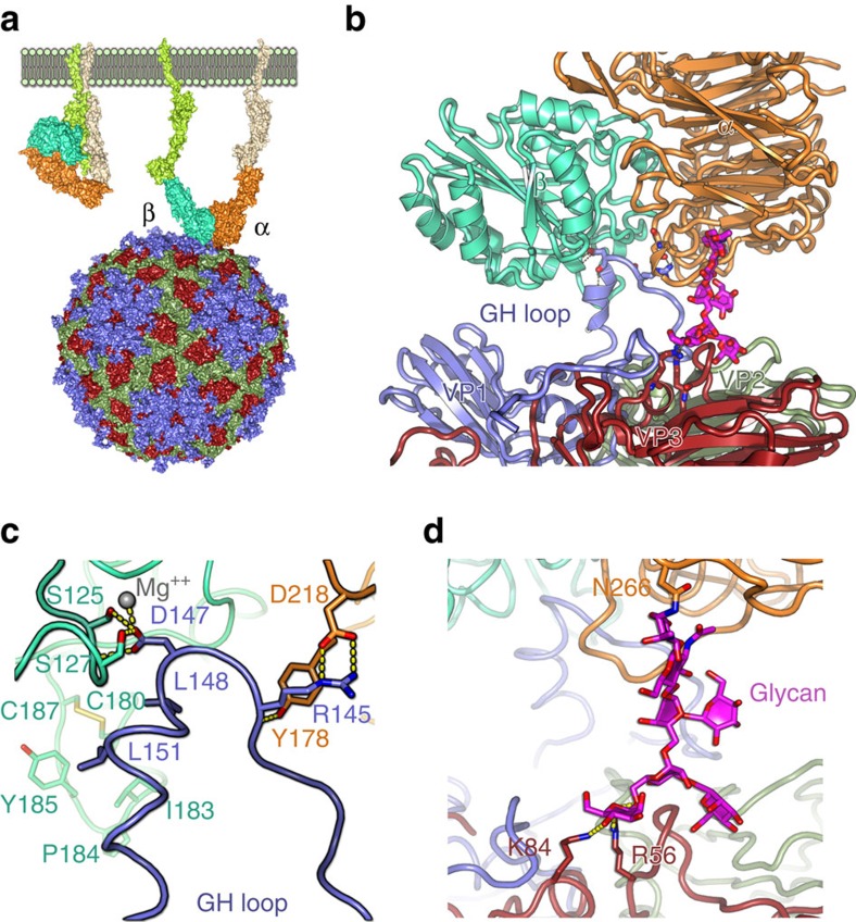
Foot-and-mouth disease virus (FMDV) mediates cell entry by attachment to an integrin receptor, generally αvβ6, via a conserved arginine-glycine-aspartic acid (RGD) motif in the exposed, antigenic, GH loop of capsid protein VP1. Infection can also occur in tissue culture adapted virus in the absence of integrin via acquired basic mutations interacting with heparin sulphate (HS); this virus is attenuated in natural infections. HS interaction has been visualized at a conserved site in two serotypes suggesting a propensity for sulfated-sugar binding. Here we determined the interaction between αvβ6 and two tissue culture adapted FMDV strains by cryo-electron microscopy. In the preferred mode of engagement, the fully open form of the integrin, hitherto unseen at high resolution, attaches to an extended GH loop via interactions with the RGD motif plus downstream hydrophobic residues. In addition, an N-linked sugar of the integrin attaches to the previously identified HS binding site, suggesting a functional role.


Conflict of interest statement

The authors declare no competing financial interests.

Figures

Figure 1
Figure 1. Apo and holo αvβ6-FMDV complexes.
(a) The density for the αvβ6-PanAsia complex determined by cryo-EM. On the left of the image the front half of the density has been cut away so that a cross-section of the binding can be seen. Depth-cueing is used such that colour indicates radius (<110 Å: cyan; 120–140 Å: yellow; >150 Å red). Clouds of red density show the bound integrin. (b) EM structure of αvβ6-O1M, depicted as for a. (c) The structure of PanAsia determined by crystallography. Colouring reflects the different viral proteins, VP1 (blue), VP2 (green) and VP3 (red). One asymmetric unit is outlined. (d) Enlargement of one asymmetric unit coloured as in c. Two conformations of the VP1 GH loop are rendered using a ribbon representation and the RGD motif is depicted with spheres. The conformational change from that seen in reduced O1 virus (PDB:1FOD) (blue) to the integrin bound conformation (cyan) is depicted with an arrow. Some of the residues involved in heparin sulfate (HS) binding are shown as orange spheres and the binding site is labelled. In all panels, one 2-fold, 3-fold and 5-fold axis of icosahedral symmetry has been labelled by a black ellipse, triangle and pentagon, respectively, and all panels show the same view down a 2-fold axis of symmetry.
Figure 2
Figure 2. αvβ6-FMDV binding modes.
Two predominant binding modes are shown for PanAsia (a,b) and for O1M (c,d). The left panel shows a view down onto the capsid surface (just a pentamer of each virus is shown colour-coded as in Fig. 1c). The integrin is drawn in cartoon representation with the alpha-helices rendered as rods; αv is orange and β6 is green. The right hand panels depict orthogonal side-views of the complexes. The integrin is drawn as in the left panel interacting with a protomer of the virus in cartoon style and colour coded as in Fig. 1c with VP4 in yellow. The FMDV VP1 GH loop, in blue, can be seen interacting with the integrin. The αv N-linked sugar, which forms an additional attachment to the virus, is drawn in magenta (marked with an arrow in a). The similarity in the binding mode ‘A' between the two viruses is evident.
Figure 3
Figure 3. αvβ6-FMDV binding modes.
(ad) The two predominant binding modes are shown for O PanAsia (a,b) and for O1M (c,d) as depicted in Fig. 2 but enlarged to show the electron density for the integrin component.
Figure 4
Figure 4. Close-up of mode A binding of αvβ6 to FMDV.
(a) overview in wall-eye stereo of the binding to O PanAsia (be) close-ups of the GH loops and N-linked carbohydrate density are shown for O PanAsia (b,c) and for O1M (d,e).
Figure 5
Figure 5. The major binding mode.
(a) The schematic shows the change in conformation of the integrin from the ‘closed' state (left) to the ‘open' (right) as visualized in the capsid interaction. (b) Side-view showing an FMDV biological protomer drawn in cartoon format with the proteins coloured by chain as in Fig. 1c. The integrin subunits are drawn in ribbon representation and coloured as in Fig. 2. Key interacting residues are drawn as sticks. The sugar is drawn as sticks in magenta. (c) Close-up of the VP1 GH loop/integrin interactions shown in a with the protein backbone drawn as a ribbon and the interacting side-chains as sticks. The magnesium ion expected to coordinate the MIDAS interaction is included as a grey sphere. (d) Close-up of the sugar interactions shown in a with the proteins depicted as in b.

References

    1. Knipe D. M. & Howley P. M. Fields' Virology Lippincott Williams & Wilkins (2007).
    1. Knowles N. J. & Samuel A. R. Molecular epidemiology of foot-and-mouth disease virus. Virus Res. 91, 65–80 (2003). - PubMed
    1. Acharya R. et al. The three-dimensional structure of foot-and-mouth disease virus at 2.9 A resolution. Nature 337, 709–716 (1989). - PubMed
    1. Lea S. et al. The structure and antigenicity of a type C foot-and-mouth disease virus. Structure 2, 123–139 (1994). - PubMed
    1. Curry S. et al. Perturbations in the surface structure of A22 Iraq foot-and-mouth disease virus accompanying coupled changes in host cell specificity and antigenicity. Structure 4, 135–145 (1996). - PubMed

Publication types

LinkOut - more resources