. 2017 May 23:8:15557.
doi: 10.1038/ncomms15557.

Exercise induces cerebral VEGF and angiogenesis via the lactate receptor HCAR1

Affiliations

Exercise induces cerebral VEGF and angiogenesis via the lactate receptor HCAR1

Cecilie Morland et al. Nat Commun. .

Abstract

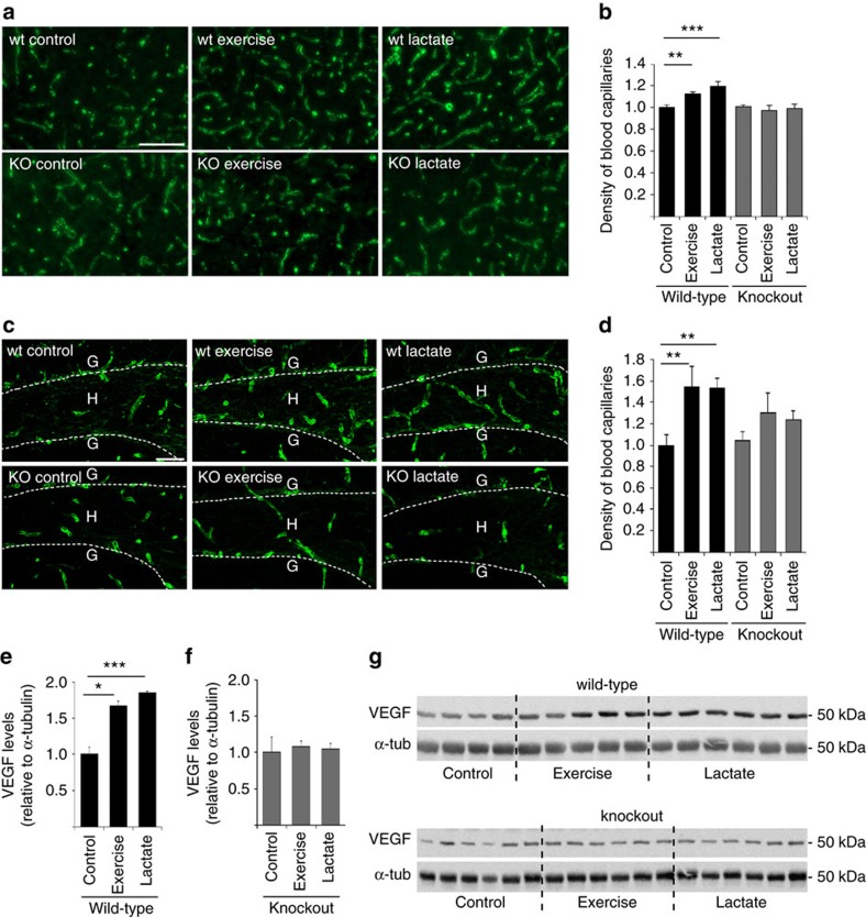
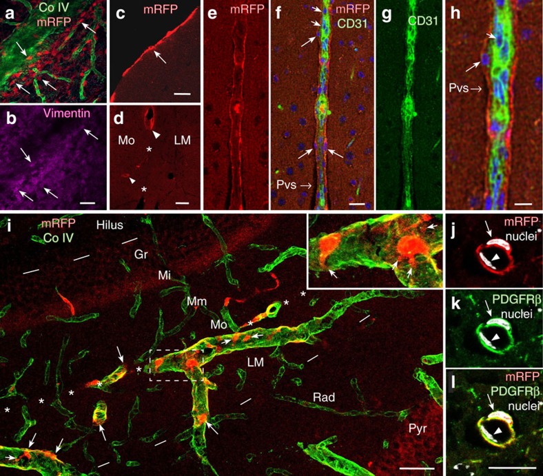
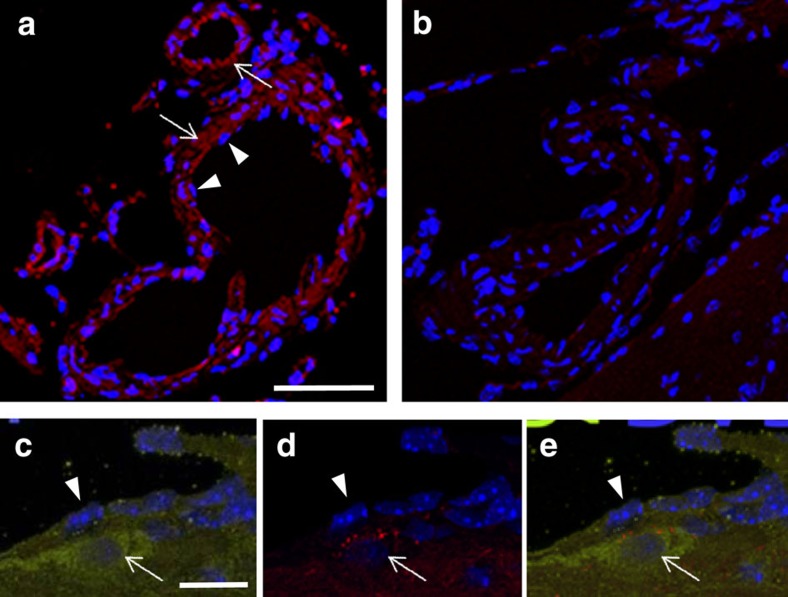
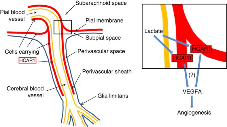
Physical exercise can improve brain function and delay neurodegeneration; however, the initial signal from muscle to brain is unknown. Here we show that the lactate receptor (HCAR1) is highly enriched in pial fibroblast-like cells that line the vessels supplying blood to the brain, and in pericyte-like cells along intracerebral microvessels. Activation of HCAR1 enhances cerebral vascular endothelial growth factor A (VEGFA) and cerebral angiogenesis. High-intensity interval exercise (5 days weekly for 7 weeks), as well as L-lactate subcutaneous injection that leads to an increase in blood lactate levels similar to exercise, increases brain VEGFA protein and capillary density in wild-type mice, but not in knockout mice lacking HCAR1. In contrast, skeletal muscle shows no vascular HCAR1 expression and no HCAR1-dependent change in vascularization induced by exercise or lactate. Thus, we demonstrate that a substance released by exercising skeletal muscle induces supportive effects in brain through an identified receptor.


Conflict of interest statement

The authors declare no competing financial interests.

Figures

Figure 1
Figure 1. HCAR1 regulates VEGFA and capillary density in response to exercise.
(a) Collagen IV-labelled capillaries in the sensorimotor cortex grey matter of wild-type or Hcar1 knockout mice exposed to vehicle injections (control), treadmill exercise or lactate injections, 5 days a week for 7 consecutive weeks. Scale bar, 100 μm. (b) Capillary density (per cent of the total area, normalized to wild-type control) in the sensorimotor cortex. Mean±s.e.m. of n=7 wild-type controls, seven wild-type exercise, six wild-type lactate, five knockout controls, four knockout exercise and six knockout lactate mice. Analysis of variance (ANOVA), P=0.001; Fisher's least significant difference (LSD) post hoc test, **P<0.01; ***P<0.001. (c) Collagen IV-labelled capillaries in the dentate gyrus (DG) of the hippocampus of wild-type or Hcar1 knockout mice treated as in a. Stippled line, the inner border of the granule cell layer (G), circumscribing the sampled area, hilus (H). Scale bar, 50 μm. (d) Capillary density (see b) in the hilus. Mean±s.e.m. of n mice (n=4–7, as specified below for diameters). ANOVA, P=0.022; Fisher's LSD post hoc test, **P<0.01. Capillary external diameters (μm) in the same areas were unchanged (mean±s.e.m. (n)): wild-type control 5.8±0.2 (6), wild-type exercise 5.7±0.1 (7), wild-type lactate 5.7±0.2 (6), knockout control 5.8±0.3 (5), knockout exercise 6.0±0.1 (4), knockout lactate 5.8±0.2 (6). ANOVA, P=0.93. (e) Quantification of VEGFA in hippocampus of wild-type animals. Data are relative to α-tubulin (α-tub), normalized to wild-type control, mean±s.e.m. of n=4 wild-type control mice, five wild-type exercised mice and six mice treated with lactate, ANOVA, P=0.001; Dunnetts's T3 post hoc test, *P<0.05, ***P<0.001. (f) Quantification of VEGFA in hippocampus of knockout animals presented as in e. n=6 mice per group. ANOVA, P=0.88. (g) Western blots of VEGFA underlying e,f (uncropped scans shown in Supplementary Fig. 6a).
Figure 2
Figure 2. Survey of HCAR1 in pia mater and at pial blood vessels supplying the brain.
(a,b) Surface views of the brain of a mRFP-HCAR1 reporter mouse. (a) Right arteria cerebri media (Mca), lateral view. Scale bar, 900 μm. (b) Branch of left Mca, top view, and vein draining into the superior sinus sagittalis (Sss). Branches penetrating the brain parenchyma are seen as black holes in b (red arrowheads). mRFP fluorescence appears enriched along blood vessels. Scale bar, 300 μm. (c,d) Two-photon imaging of pia mater in a live and walking mRFP-HCAR1 reporter mouse showing leptomeningeal cells (arrows) in the vicinity of two pial blood vessels (approximate vessel positions indicated by red dotted lines). (c) Stack of superimposed optical sections. Scale bars, 100 μm. (d) Single optical section (1 μm); frame shown is magnified as inset in c; stippled frame in c indicates) position of frame in stack. (e) Surface view of the cortex from mRFP-HCAR1 reporter mouse showing fluorescent pial vessel outline (arrowheads) and lumen (green) after retrograde injection of FITC-dextran agarose in the v. jugularis. Scale bar, 300 μm. Inset: magnified view showing space between green and red corresponding to vessel wall (small arrows).
Figure 3
Figure 3. Details of HCAR1-expressing cells in pia mater and pial and brain vessels.
(a,b) Pial vessel with emerging capillaries labelled for basement membrane collagen IV (CoIV, green), decorated by leptomeningeal cells that contain mRFP-HCAR1 (red) and co-localize fibroblast marker vimentin (magenta, b); confocal images from top frozen section tangential to the brain surface. Scale bar, 70 μm. (c) Pial cells (arrow) in confocal image of parasagittal frozen section perpendicular to the cerebral cortex of mRFP-HCAR1 reporter mouse. Scale bar, 20 μm. (d) In the extension of pia in fissura hippocampi (*), which separates hippocampus stratum lacunosum-moleculare (LM) from the molecular layer of area dentata (Mo), mRFP-HCAR1 is in select blood vessels (arrowheads), including ones (small arrowhead) penetrating into the Mo. Scale bar, 50 μm. (eh) Blood vessel penetrating into the cerebral cortex with a sheath of mRFP-HCAR1-containing perivascular cells (long arrows), CD31 in endothelial cells (short arrows, green; fg) and DAPI-stained cell nuclei (blue; f); single optical section (1.38 μm). The picture is compatible with a slight mRFP-HCAR1 signal also in endothelial cells. Subpial/paravascular/perivascular space (Pvs). Scale bar, 20 μm (eg). (h) Magnification of part of f. Scale bar, 10 μm. (i) mRFP-HCAR1 in the hippocampus, magnified frame showing details of mRFP-HCAR1-carrying cells (arrows, red), which extend processes (small arrows) around blood vessels (CoIV-labelled, green) that penetrate into the hippocampus through the extension of pia in the fissura hippocampi (indicated by white asterisks, brain surface to the left outside the picture). Hil, hilus of area dentata; Gr, granule layer of area dentata; Mi, Mm, Mo, inner, middle and outer zones of the molecular layer of area dentata; LM, Rad and Pyr, lacunosum-moleculare, radiatum and pyramidal layers of hippocampus CA1. Dashes mark borders between Hil and Gr, and between LM and Rad. Scale bar, 50 μm. (jl) Small vessel in the cerebral neocortex, surrounded by cell co-expressing mRFP-HCAR1 (j, red; l, yellow) and the pericyte-associated protein PDGFRβ (k, green; l, yellow). Staining of nuclei (arrow and arrowhead, DAPI, white) reveals that an endothelial cell (arrowhead) is located between the lumen and the mRFP-HCAR1/PDGFRβ-co-expressing cell. Scale bar, 15 μm.
Figure 4
Figure 4. HCAR1 immunoreactivity colocalizes with mRFP-HCAR1 at pial vessels.
(a,b) HCAR1 immunoreactivity (red) is in the wall of blood vessels in pia mater (arrows), in wild-type (a), but not in knockout (b). Nuclei of endothelial cells (arrowheads) are on the luminal side of the immunoreactivity (a). Scale bar, 30 μm. (ce) HCAR1 immunoreactivity (green, c,e) is highly expressed in a leptomeningeal cell (arrow) on the abluminal side of the endothelium (arrowhead), which shows a slight HCAR1 immunoreactivity. In this reporter mouse, immunoreactivity to mRFP-HCAR1 (red, d,e) is seen in cytoplasmic granules in the leptomeningeal cell. Nuclei are blue (DAPI). All sections are from paraffin-embedded brain tissue and exposed to heat-induced epitope retrieval. Scale bars, (a,b), 30 μm; (ce), 10 μm.
Figure 5
Figure 5. Organization of cells that carry HCAR1 and of the angiogenic action of lactate.
Blood-borne lactate from exercising muscle penetrates the blood vessel wall (yellow) through monocarboxylate transporters located in the vascular endothelium (which represents the blood–brain barrier). Extravascular lactate (from blood or generated in the brain parenchyma upon neural activation) is freely diffusible in the perivascular/subpial space, thereby bathing the leptomeningeal fibroblast-like cells carrying HCAR1 (red). Magnified inset indicates possible, yet unidentified (?), pathways leading from activation of HCAR1 in the cells in pia and perivascular sheaths to increased VEGFA and subsequent enhanced angiogenesis. The perivascular sheath extends as separate HCAR1-expressing pericyte-like cells at intracerebral microvessels, which may also contribute in the angiogenic process. Although apparently devoid of mRFP-HCAR1, other cells may possibly express low levels of HCAR1. HCAR1 may stimulate VEGFA in the same cells, or in other cells, through mediators. In addition to its angiogenic action, VEGFA has neurotrophic effects. Importantly, all blood to the brain parenchyma has to pass in close proximity to the perivascular sheath of HCAR1-carrying cells and therefore can convey products released from these cells upon activation of the receptor; blood to the hippocampus passes through vessels (such as the ones shown in Fig. 3i) entering in the hippocampal fissure, an extension of the pia mater that penetrates deep into the centre of the hippocampal formation. (The anatomical sketch is based on ref. 54).

References

    1. van Praag H., Shubert T., Zhao C. & Gage F. H. Exercise enhances learning and hippocampal neurogenesis in aged mice. J. Neurosci. 25, 8680–8685 (2005). - PMC - PubMed
    1. Barnes J. N. Exercise, cognitive function, and aging. Adv. Physiol. Educ. 39, 55–62 (2015). - PMC - PubMed
    1. Paillard T. Preventive effects of regular physical exercise against cognitive decline and the risk of dementia with age advancement. Sports Med. Open 1, 4 (2015). - PMC - PubMed
    1. Ding Y. H. et al.. Cerebral angiogenesis and expression of angiogenic factors in aging rats after exercise. Curr. Neurovasc. Res. 3, 15–23 (2006). - PubMed
    1. Wightman E. L. et al.. Dietary nitrate modulates cerebral blood flow parameters and cognitive performance in humans: a double-blind, placebo-controlled, crossover investigation. Physiol. Behav. 149, 149–158 (2015). - PubMed

Publication types

MeSH terms

Substances