. 2017 May 23:8:15216.
doi: 10.1038/ncomms15216.

Crystal structure of the receptor binding domain of the spike glycoprotein of human betacoronavirus HKU1

Affiliations

Crystal structure of the receptor binding domain of the spike glycoprotein of human betacoronavirus HKU1

Xiuyuan Ou et al. Nat Commun. .

Abstract

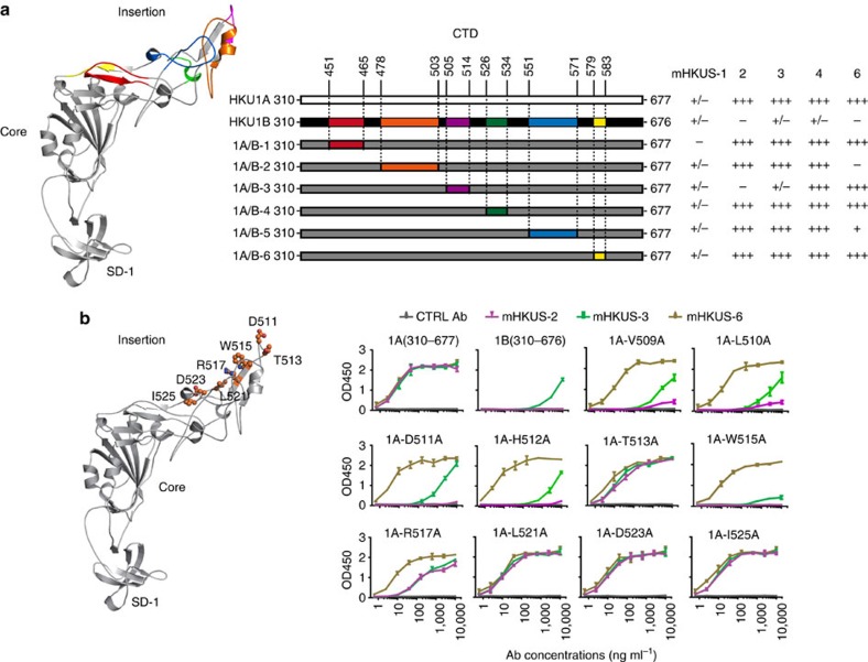
Human coronavirus (CoV) HKU1 is a pathogen causing acute respiratory illnesses and so far little is known about its biology. HKU1 virus uses its S1 subunit C-terminal domain (CTD) and not the N-terminal domain like other lineage A β-CoVs to bind to its yet unknown human receptor. Here we present the crystal structure of HKU1 CTD at 1.9 Å resolution. The structure consists of three subdomains: core, insertion and subdomain-1 (SD-1). While the structure of the core and SD-1 subdomains of HKU1 are highly similar to those of other β-CoVs, the insertion subdomain adopts a novel fold, which is largely invisible in the cryo-EM structure of the HKU1 S trimer. We identify five residues in the insertion subdomain that are critical for binding of neutralizing antibodies and two residues essential for receptor binding. Our study contributes to a better understanding of entry, immunity and evolution of CoV S proteins.


Conflict of interest statement

The authors declare no competing financial interests.

Figures

Figure 1
Figure 1. Inhibition of HKU1 virus infection by 1A-S310-677aa.
(a) Schematic diagram of HKU1 S protein, modified from Kirchdoerfer et al. NTD, N-terminal domain; CTD, C-terminal domain; SD-1, subdomain-1; SD-2, subdomain-2; RBD, receptor-binding domain; FP, fusion peptide; HR-N, N-terminal heptad repeat; HR-C, C-terminal heptad repeat; TMD, transmembrane domain; cleavage site, furin cleavage site between S1 and S2. The amino acid numbers are from the S protein of genotype A HKU1 virus. (b) ELISA was performed using purified 1A-S310-677aa and purified antibodies. The experiments were done in triplicate and the s.d.‘s (n=3) were shown as error bar. The experiments were performed twice and one representative is shown. Differentiated HTBE cells were incubated with 20 μM of 1A-310-677aa protein at 37 °C for 1 h. HKU1 viruses were diluted into the same amount of proteins and added onto HTBE cells for 4 h. Forty-eight hours after inoculation, cells were fixed and stained with polyclonal rabbit anti HKU1 S antibodies 1814 at a dilution at 1:100 (c), and released viruses at 24 h post inoculation from apical wash were analysed using real-time PCR (d). The amount of HKU1 viral RNA from BSA control was set as 100%. Scale bar, 50 μm. The experiments were done in triplicate and the s.d.‘s (n=3) were shown as error bar. The experiments were performed twice and one representative is shown.
Figure 2
Figure 2. Crystal structure of 1A-310-677aa.
(a, top) Ribbon model of 1A-310-677aa; bottom: schematic diagram of 1A-310-677aa. The core subdomain is shown in blue, the insertion loop is shown in magenta, and SD-1 subdomain is shown in green. Twelve pairs of disulfite bridges are numbered and shown in yellow and stick model. The amino acid numbers are from the S protein of the genotype A HKU1 virus. (b) Schematic illustration of the topological graph of 1A- 310-677aa. β-strands are shown with brown arrows, α-helices are shown with bright blue cylinders.
Figure 3
Figure 3. Structural comparison of CTDs of different β-CoV S proteins.
The core subdomains are blue, and the insertion loops are magenta. (a) HKU1 CTD; (b) MHV CTD (PDB: 3JCL); (c) (PDB: 2AJF); (d) MERS-CoV RBD (PDB: 4L72); (e) HKU4 RBD (PDB: 4QZV) and (f) HKU9 RBD (PDB: 5GYQ).
Figure 4
Figure 4. Integrated model of HKU1 S protein trimer with the crystal structure of 1A-S310-677aa.
(a) The trimeric model of HKU1 S protein with the crystal structure of 1A-S310-677aa. (left) side view; (right) top view. The model is built by superimposing the crystal structure of 1A-S310-677aa into the Cryo-EM structure of the HKU1 S protein trimer based on the structure alignment of the core subdomain. The cryo-EM structure of the trimeric HKU1 S protein is shown in grey. The three crystal structures of 1A-S310-677aa are labelled in blue, magenta and green, respectively. The red spheres are sugar moieties found in the crystal structure of 1A-S310-677aa. (b) Superimposition of 1A-S310-677aa onto the cryo-EM density of the HKU1 S protein. The crystal structure of 1A-S310-677aa is shown in blue and the cryo-EM density map is shown in orange. N-linked glycan moieties are shown in red stick.
Figure 5
Figure 5. Epitope mapping for neutralizing mHKUS-2 and mHKUS-3 antibodies in 1A-S310-677aa.
(a, left) Structure of 1A-S310-677aa; middle, schematic diagram of different 1A/B CTD chimeras; right, binding of different mHKUS antibodies to 1A/B chimeric proteins measured by ELISA. +++, positive signal with antibody concentration <10 ng ml−1 ++, positive signal with antibody concentration 10–100 ng ml−1; +, positive signal with antibody concentration 100–1,000 ng ml−1; +/−, positive signal with antibody concentration >1,000 ng ml−1; -, no signal with antibody concentration at 10 μg ml−1. The amino acid residue numbers are from S protein of genotype A HKU1 virus. Experiments were done twice. (b) ELISAs were performed using purified proteins and purified antibodies. The experiments were done in triplicate and the s.d.‘s (n=3) were shown as error bar. The experiments were performed twice and one representative is shown.
Figure 6
Figure 6. Inhibition of HKU1 virus entry by mutant 1A-S310-677aa proteins.
(a) The purified mutant proteins were assayed for their affinity to monoclonal antibodies mHKUS-2, mHKUS-4, and mHKUS-6 by ELISA. Differentiated HTBE cells were incubated with 20 μM of 1A-S310-677aa, 1B-S310-676aaor mutant CTD proteins at 37 °C for 1 h. HKU1 viruses were diluted into the same amount of proteins and added onto the HTBE cells for 4 h. The released viruses from apical wash at 24 h post inoculation were analysed using real-time PCR (b), and cells were fixed and stained with polyclonal rabbit anti HKU1 S antibodies 1814 at a dilution of 1:100 at 48 h post inoculation (c). Scale bar, 50 μm. The amount of HKU1 viral RNA in the BSA control was set as 100%. The experiments were done in triplicate, and the s.d.‘s (n=3) were shown as error bar. The experiments were performed twice and one representative is shown.

References

    1. Masters P. S. & Perlman S. in Fields Virology, 6th edn, Vol. 1 (eds Knipe, D. M. & Howley, P. M.) 825–858 (Wolters Kluwer Health/Lippincott Williams & Wilkins, 2013).
    1. Viruses, I.C.o.T.o. Virus Taxonomy: 2011 Release. Available at: http://ictvonline.org/virusTaxonomy.asp?version=2011 (2011).
    1. Drosten C. et al. Identification of a novel coronavirus in patients with severe acute respiratory syndrome. N. Engl. J. Med. 348, 1967–1976 (2003). - PubMed
    1. Ksiazek T. G. et al. A novel coronavirus associated with severe acute respiratory syndrome. N. Engl. J. Med. 348, 1953–1966 (2003). - PubMed
    1. Peiris J. S. et al. Coronavirus as a possible cause of severe acute respiratory syndrome. Lancet 361, 1319–1325 (2003). - PMC - PubMed

Publication types

MeSH terms