. 2017 May 19;9(5):173.
doi: 10.3390/toxins9050173.

RBC Adherence of Immune Complexes Containing Botulinum Toxin Improves Neutralization and Macrophage Uptake

Affiliations

RBC Adherence of Immune Complexes Containing Botulinum Toxin Improves Neutralization and Macrophage Uptake

Fetweh H Al-Saleem et al. Toxins (Basel). .

Abstract

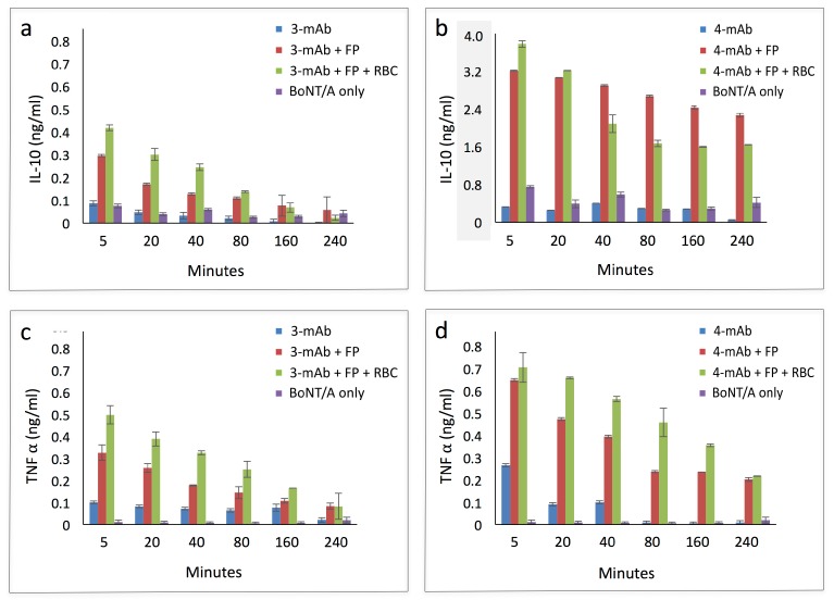
In the paralytic disease botulism, the botulinum neurotoxin (BoNT) passes through the bloodstream to reach and inactivate neuromuscular junctions. Monoclonal antibodies (mAbs) may be useful BoNT countermeasures, as mAb combinations can rapidly clear BoNT from the blood circulation. We have previously shown that the BoNT-neutralizing potency of mAbs can be improved through red blood cell (RBC) immunoadherence. For example, a fusion protein (FP) that adheres biotinylated mAbs to the RBC surface enabled a pair of mAbs to neutralize 5000 LD50 BoNT/A in the mouse protection assay. Here, we added two mAbs to that combination, creating a 4-mAb:FP complex that neutralized 40,000 LD50 BoNT/A in vivo, and analyzed functional correlates of neutralization. The FP enhanced potency of BoNT/A immune complexes, providing the greatest magnitude of benefit to the 4-mAb combination. RBC binding of a BoNT/A complexed with 4-mAb:FP exhibited a bi-phasic clearance process in vivo. Most of the complexes were cleared within five minutes; the rest were cleared gradually over many hours. Peritoneal macrophages showed better uptake of the 4-mAb complex than the 3-mAb complex, and this was not affected by the presence of the FP. However, the addition of RBCs to the 4-mAb:FP BoNT/A doubled macrophage uptake of the complexes. Lastly, the 4-mAb:FP BoNT/A complex synergistically induced M2 macrophage polarization, as indicated by IL-10 expression, whether or not RBCs were present. RBC-targeted immunoadherence through the FP is a potent enhancer of mAb-mediated BoNT/A neutralization in vivo, and can have positive effects on BoNT/A sequestration, immune complex uptake, and macrophage activation.

Keywords: biodistribution; botulinum toxin; clearance; immune complex; macrophage activation; mechanism; monoclonal antibody; neutralization; pharmacokinetic; red blood cell.


Conflict of interest statement

The authors declare no conflict of interest. The funding sponsors had no role in the design of the study; in the collection, analyses, or interpretation of data; in the writing of the manuscript, and in the decision to publish the results.

Figures

Figure 1
Figure 1
Competitive binding assays. Binding of biotinylated 6A or 3B3 mAbs to serial dilutions of RI-BoNT/A was assessed in the absence of potential competitor mAbs. Assays tested inhibition of biotinylated 6A binding in the presence of un-modified mAbs: CR2 (A), 3B3 (B), 15A (C) and 4LCA (D), or the inhibition of biotinylated 3B3 binding in the presence of CR2 (E) or 3B3 (F). Percent reduction in binding is shown in the top left of each panel and was calculated using the trapezoidal method.
Figure 2
Figure 2
Binding 3-mAb:FP and 4-mAb:FP immune complexes to RBCs in vitro. Washed Swiss Webster RBCs were incubated with RI-BoNT/A bound to 3-mAb:FP (left) or 4-mAb:FP (right). Complex binding to RBCs was detected using an APC-conjugated anti-human IgG and flow cytometry. Tracings include RBCs only (red), RBCs with the APC anti-human IgG (blue), and RBCs with the BoNT/A complexes and the APC anti-human IgG (orange).
Figure 3
Figure 3
Qualitative assessment of BoNT/A in solution before and after incubation with murine RBCs. RI-BoNT/A contained in the 3-mAbs:FP complex was detected by ELISA in a solution before (Pre) and after (Post) its incubation with the RBCs, as shown in Figure 2, and compared to a buffer-only sample (Blank). Error bars indicate the S.E.M.
Figure 4
Figure 4
Time course of in vivo RBC adherence of BoNT/A complexed with 4-mAb:FP. The 4-mAb:FP complex bound to 200 ng BoNT/A (40,000 LD50) was injected i.v. into groups of five Swiss Webster mice, and their RBCs were tested for complex binding with an the APC anti-human IgG and flow cytometry. Blood samples were tested at 5, 20, 40, 80, 160 and 240 min, and the data for each group were averaged. For the zero timepoint, complexes were incubated with RBCs in vitro. (A) Flow cytometry tracings. (B) Calculated Mean Fluorescence Intensity (MFI) of the curves shown in Figure 4a. The zero timepoint value is depicted in light coloring to emphasize that it is not an in vivo sample. Error bars in Figure 4b indicate the S.D.
Figure 4
Figure 4
Time course of in vivo RBC adherence of BoNT/A complexed with 4-mAb:FP. The 4-mAb:FP complex bound to 200 ng BoNT/A (40,000 LD50) was injected i.v. into groups of five Swiss Webster mice, and their RBCs were tested for complex binding with an the APC anti-human IgG and flow cytometry. Blood samples were tested at 5, 20, 40, 80, 160 and 240 min, and the data for each group were averaged. For the zero timepoint, complexes were incubated with RBCs in vitro. (A) Flow cytometry tracings. (B) Calculated Mean Fluorescence Intensity (MFI) of the curves shown in Figure 4a. The zero timepoint value is depicted in light coloring to emphasize that it is not an in vivo sample. Error bars in Figure 4b indicate the S.D.
Figure 5
Figure 5
Detection of BoNT/A in plasma over time. In an experiment similar to the one shown in Figure 4, the 4-mAb:FP complex, bound to 200 ng BoNT/A (40,000 LD50), was injected i.v. into Swiss Webster mice. RI-BoNT/A in plasma was tested by ELISA at 5, 20, 40, 80, and 160 min in groups with 5 mice each. Control: dilution buffer only. Error bars indicate the S.E.M.
Figure 6
Figure 6
Phagocytosis of BoNT/A immune complexes by peritoneal macrophages. Alexa Fluor 488-labeled RI-BoNT/A was bound to the 3-mAb or 4-mAb combinations, alone or in combination with the FP or with FP and RBCs. Panel (a) RI-BoNT/A alone. Panels (bf) RI-BoNT/A with: (b) 3-mAb, (c) 3-mAb + FP, (d) 4-mAb, (e) 4-mAb + FP, (f) 4-mAb + FP + RBCs.
Figure 7
Figure 7
Quantitation of the uptake of RI-BoNT/A by macrophages in the experiment shown in Figure 6. Internalized Alexa-Fluor labeled RI-BoNT/A was assessed by measuring individual cells using confocal microscopy and IMAGEj software. The mean corrected total cell intensity (CTCF) is shown. Error bars indicate the S.E.M.
Figure 8
Figure 8
Cytokine production by peritoneal macrophages in the presence of RI-BoNT/A immune complexes and RBCs. Peritoneal macrophages were cultured in vitro and incubated with RI-BoNT/A bound to the 3-mAb or 4-mAb combinations, alone or in combination with the FP or with the FP and RBCs. The production of IL-10 and TNF-α was measured over time using a luminescence capture assay. Top panels: IL-10 production induced by complexes containing the (a) 3-mAb or (b) 4-mAb. Bottom panels: TNF-α production induced by complexes containing the (c) 3-mAb or (d) 4-mAb. The y-axis in panel (b) is greyed to emphasize that it has a different scale than panels (a,c,d).

References

    1. Arnon S.S., Schechter R., Inglesby T.V., Henderson D.A., Bartlett J.G., Ascher M.S., Eitzen E., Fine A.D., Hauer J., Layton M., et al. Botulinum toxin as a biological weapon: Medical and public health management. JAMA. 2001;285:1059–1070. doi: 10.1001/jama.285.8.1059. - DOI - PubMed
    1. Simpson L. The life history of a botulinum toxin molecule. Toxicon. 2013;68:40–59. doi: 10.1016/j.toxicon.2013.02.014. - DOI - PubMed
    1. Al-Saleem F.H., Ancharski D.M., Ravichandran E., Joshi S.G., Singh A.K., Gong Y., Simpson L.L. The role of systemic handling in the pathophysiologic actions of botulinum toxin. J. Pharmacol. Exp. Ther. 2008;326:856–863. doi: 10.1124/jpet.108.136242. - DOI - PubMed
    1. Nowakowski A., Wang C., Powers D.B., Amersdorfer P., Smith T.J., Montgomery V.A., Sheridan R., Blake R., Smith L.A., Marks J.D. Potent neutralization of botulinum neurotoxin by recombinant oligoclonal antibody. Proc. Natl. Acad. Sci. USA. 2002;99:11346–11350. doi: 10.1073/pnas.172229899. - DOI - PMC - PubMed
    1. Sepulveda J., Mukherjee J., Tzipori S., Simpson L.L., Shoemaker C.B. Efficient serum clearance of botulinum neurotoxin achieved using a pool of small antitoxin binding agents. Infect. Immun. 2010;78:756–763. doi: 10.1128/IAI.01084-09. - DOI - PMC - PubMed

Publication types

MeSH terms