. 2017 Jul;51(1):63-74.
doi: 10.3892/ijo.2017.4002. Epub 2017 May 17.

Metadherin regulates actin cytoskeletal remodeling and enhances human gastric cancer metastasis via epithelial-mesenchymal transition

Affiliations

Metadherin regulates actin cytoskeletal remodeling and enhances human gastric cancer metastasis via epithelial-mesenchymal transition

Yaqiong Du et al. Int J Oncol. 2017 Jul.

Abstract

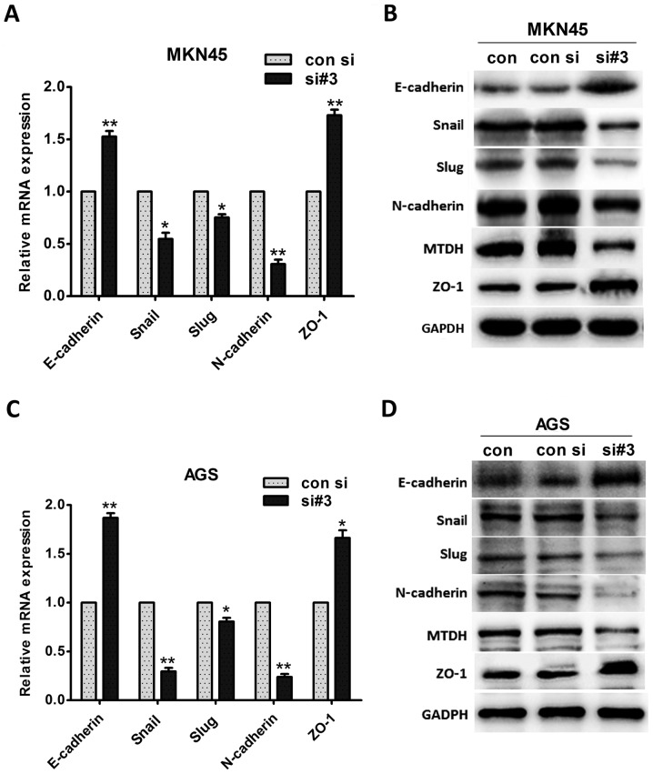
Metadherin (MTDH) can be recruited to mature tight junction complexes, and it regulates mesenchymal marker protein expression in many tumors and promote cancer metastasis. This study investigated the influence of MTDH expression on gastric cancer and to elucidate the potential mechanisms by which MTDH regulates actin cytoskeletal remodeling and enhances human gastric cancer metastasis via epithelial-mesenchymal transition (EMT). Relative MTDH mRNA expression levels were assessed by quantitative real-time PCR (Q-PCR), and MTDH protein expression levels and localization were evaluated via immunohistochemical (ICH) staining. We studied the role of MTDH in cancer cell migration and invasion by modulating MTDH expression in the gastric cancer cell lines MKN45 and AGS. We also confirmed the functions of MTDH through in vivo experiments. We found that MTDH expression levels were correlated with lymph node metastasis, TNM stages and decreased OS (P=0.002, <0.001 and 0.010, respectively) in human gastric cancer and that MTDH upregulation promoted EMT in vitro. Consistent with this finding, MTDH downregulation inhibited cell migration and invasion in vitro and suppressed tumor growth and metastasis in vivo. Furthermore, MTDH knockdown regulated actin cytoskeletal remodeling and inhibited EMT. Overall, our results provide a novel role for MTDH in regulating gastric cancer metastasis.


Figures

Figure 1
Figure 1
MTDH expression is upregulated in gastric cancer and is positively correlated with gastric cancer metastasis ability. (A) qPCR analysis of the expression levels of MTDH mRNA in 31 fresh pairs of primary gastric cancer tissues after the expression levels were normalized against the basal expression levels of MTDH mRNA in adjacent normal gastric mucosal tissues. (B) MTDH mRNA expression levels in primary gastric cancer patients with or without lymph node metastasis (n=9 and n=22, respectively). Data are the mean ± SD. (C) Immunohistochemistry staining for MTDH expression in adjacent normal gastric mucosal tissues, primary gastric cancer tissues without metastasis, and gastric cancer tissues with metastasis. Scale bar, 100 µm. (D) The percentages of MTDH-positive cells were different among the normal, primary tumor, and metastasis groups. Data are the mean ± SD. (E). Kaplan-Meier survival curves based on MTDH expression in gastric cancer samples. Blue line: patients with high MTDH expression, median overall survival time=36 months and n=56; Purple line: patients with low MTDH expression, median overall survival time 51 months and n=27. *P<0.05, **P<0.01, ***P<0.001.
Figure 2
Figure 2
MTDH expression in gastric cancer cells and stable shMTDH gastric cancer cells. (A and B) qPCR and western blot analysis of MTDH expression levels in the gastric cancer cell lines MKN45, AGS, SGC7901, and KATO-III and the normal gastric mucosa cell line GES-1. (C and D) MKN45 and AGS cells were transfected with three MTDH-targeting siRNAs (si#1, si#2, si#3) and a negative control siRNA (con si). After 24 h, MTDH protein and mRNA expression levels were determined by western blotting and qPCR, respectively. Data are the mean ± SD. (E and F) The stable shMTDH gastric cancer cell lines, namely, the MKN45-sh#3 and AGS-sh#3 cell lines, were established, and immunofluorescence staining and western blot analysis demonstrated the interfering efficiency of the MTDH-targeting shRNA. *P<0.05, **P<0.01, ***P<0.001.
Figure 3
Figure 3
Cell invasion and migration were inhibited, and actin cytoskeletal remodeling was regulated by MTDH expression in vitro. (A and C) Scratch wound-healing assay was conducted after the MKN45 and AGS cell lines were transfected with MTDH-targeting siRNA3 (si#3) and negative control siRNA (con si) for 24 h. Observations and measurements were performed at 12-h intervals. Measurement unit, 10 µm. Scale bar, 100 µm. (B) Quantitative analysis of scratch wound-healing assay results. MKN45-si#3 and AGS-si#3 cell migration ability was decreased compared with MKN45-con si and AGS-con si cell migration ability. Data are the mean ± SD. (D and E) Transwell assay was conducted after the MKN45 and AGS cell lines were transfected with MTDH-siRNA3 (si#3) and negative control siRNA (con si) for 24 h. Normal MKN45 cells and AGS cells served as normal controls (con). MKN45-si#3 and AGS-si#3 cell migration ability was decreased compared with MKN45-con si and AGS-con si cell migration ability. Scale bar, 100 µm. Data are the mean ± SD. (F) Early cell migration and cell morphology. At 12 h, MKN45-con si cells and AGS-con si cells at the leading migration edge were spindle-shaped and were significantly elongated compared with MKN45-si#3 and AGS-si#3 cells. Scale bar, 50 µm. (G) Cell cytoimmunofluorescence assay. F-actin cytoskeletal staining. The numbers of F-actin-enriched filopodial extensions were decreased in MKN45-si#3 and AGS-si#3 cells. Scale bar, 50 µm. *P<0.05, **P<0.01, ***P<0.001.
Figure 4
Figure 4
MTDH promotes EMT in vitro. (A) qPCR analysis of the changes in EMT marker expression in MKN45 cells. Data are the mean ± SD. (B) Western blot analysis of the changes in EMT marker expression in MKN45 cells. (C) qPCR analysis of the changes in EMT marker expression in AGS cells. Data are the mean ± SD. (D) Western blot analysis of the changes in EMT marker expression in AGS cells. *P<0.05, **P<0.01, ***P<0.001.
Figure 5
Figure 5
MTDH promotes EMT via actin cytoskeletal remodeling in vitro. (A) Scratch-wound healing assay. After the MKN45 and AGS cell lines were transfected with MTDH-siRNA for 24 h, they were stimulated with TGF-β1 (5 ng/ml) for 24 h after wound placement. Scale bar, 100 µm. (B) Quantitative analysis of the relative migratory ranges of MKN45 and AGS cells. Data are the mean ± SD. Measurement unit, µm. (C) F-actin cytoskeletal staining assay. MKN45-con si and MKN45-si#3 cells were stimulated with TGF-β1 for 24 h, after which the changes in MKN45 cell morphology elicited by the treatment were assessed. Scale bar, 100 µm. (D) Western blot analysis of EMT marker protein expression in MKN45 and AGS cells after TGF-β1 stimulation for 24 h. *P<0.05, **P<0.01, ***P<0.001.
Figure 6
Figure 6
Influence of MTDH knockdown on cancer cell proliferation and metastasis in vivo. (A) Tumor proliferation in nude mice. Negative control (con sh group) and shMTDH (sh#3 group) cells derived from MKN45 cells were injected subcutaneously into the left flanks of BALB/c mice. Tumor growth was recorded, and all the mice were sacrificed after 4 weeks. (B) Measurements of all the tumor volumes and weights in the con sh and sh#3 group. Data are the mean ± SD. (C) Con sh and sh#3 cells derived from MKN45 cells were injected into the tail veins of nude mice. After 4 weeks, all the nude mice were sacrificed, and the average numbers of metastatic nodules in the lungs (shown by the black arrows) were counted. Data are the mean ± SD. (D) The metastatic tumor nodules in the lungs of nude mice were identified histologically via H&E staining. Upper panel: panoramic scans of lung tissue slices; lower panel: enlarged images, ×100. Scale bar, 200 µm. (E) Western blot analysis of MTDH, E-cadherin and N-cadherin expression in both the con sh group and the sh#3 group. (F) Immunohistochemistry was performed to determine MTDH, E-cadherin and N-cadherin expression levels in both the con sh group and the sh#3 group. *P<0.05, **P<0.01, ***P<0.001. Scale bar, 200 µm.

References

    1. Jin P, Wong CC, Mei S, He X, Qian Y, Sun L. MK-2206 co-treatment with 5-fluorouracil or doxorubicin enhances chemosensitivity and apoptosis in gastric cancer by attenuation of Akt phosphorylation. Onco Targets Ther. 2016;9:4387–4396. doi: 10.2147/OTT.S106303. - DOI - PMC - PubMed
    1. Torre LA, Bray F, Siegel RL, Ferlay J, Lortet-Tieulent J, Jemal A. Global cancer statistics, 2012. CA Cancer J Clin. 2015;65:87–108. doi: 10.3322/caac.21262. - DOI - PubMed
    1. Xia H, Chen Q, Chen Y, Ge X, Leng W, Tang Q, Ren M, Chen L, Yuan D, Zhang Y, et al. The lncRNA MALAT1 is a novel biomarker for gastric cancer metastasis. Oncotarget. 2016;7:56209–56218. - PMC - PubMed
    1. Xu W, Yang Z, Lu N. Molecular targeted therapy for the treatment of gastric cancer. J Exp Clin Cancer Res. 2016;35:1. doi: 10.1186/s13046-015-0276-9. - DOI - PMC - PubMed
    1. Brown DM, Ruoslahti E. Metadherin, a cell surface protein in breast tumors that mediates lung metastasis. Cancer Cell. 2004;5:365–374. doi: 10.1016/S1535-6108(04)00079-0. - DOI - PubMed

MeSH terms