. 2017 Jul;40(1):57-64.
doi: 10.3892/ijmm.2017.2995. Epub 2017 May 18.

MicroRNA let-7g alleviates atherosclerosis via the targeting of LOX-1 in vitro and in vivo

Affiliations

MicroRNA let-7g alleviates atherosclerosis via the targeting of LOX-1 in vitro and in vivo

Mingxin Liu et al. Int J Mol Med. 2017 Jul.

Abstract

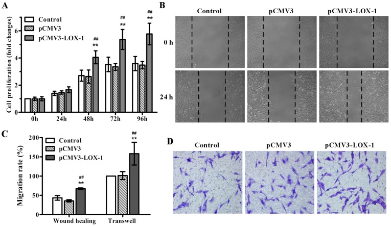
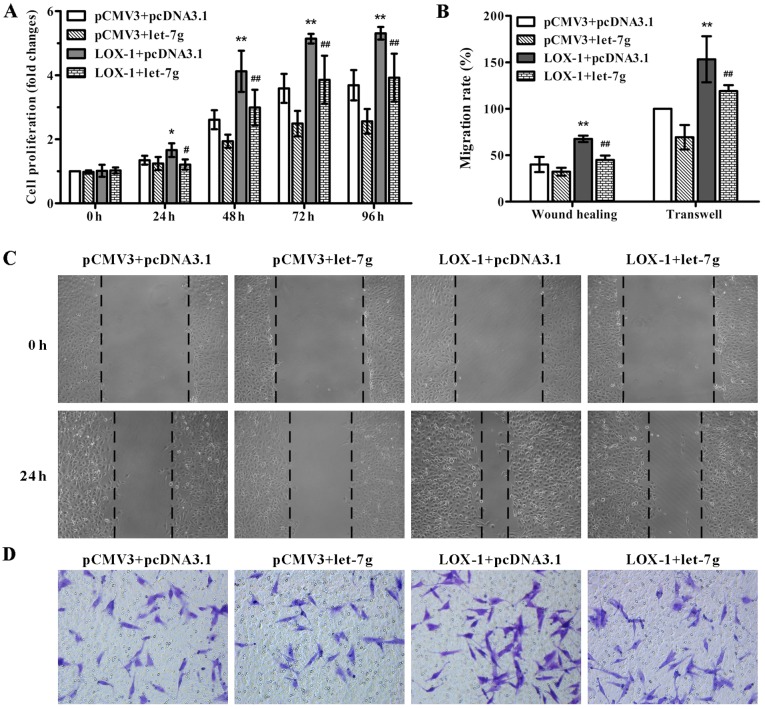
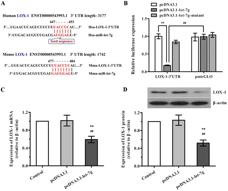
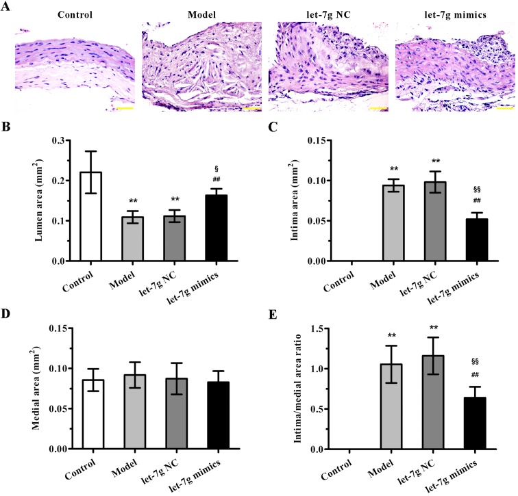
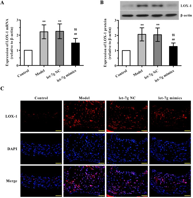
Atherosclerosis is a chronic arterial disease and the leading cause of stroke and myocardial infarction. Micro-RNAs (miRNAs or miRs) have been reported to act as essential modulators during the progression of atherosclerosis. Although miR-let-7g has been demonstrated to contribute to maintaining endothelial function and vascular homeostasis, it is not known whether miR-let-7g exerts a therapeutic effect on experimental atherosclerosis. The aim of this study was to investigate the effects of miR-let-7g on atherosclerosis in vivo and in vitro and to explore its underlying mechanisms. Data from our study showed that exogenous lectin‑like oxidized low‑density lipoprotein receptor‑1 (LOX-1 or OLR1) overexpression resulted in the significant promotion of proliferation and migration of human aortic smooth muscle cells (ASMCs), whereas such changes induced by LOX-1 were obviously suppressed by transfection of miR‑let‑7g. We later confirmed that LOX-1 is a potential target of miR-let-7g, and miR-let-7g markedly inhibited LOX-1 expression in ASMCs by directly binding to the 3' untranslated region of LOX-1. Furthermore, in a hyperlipidemic apolipoprotein E knockout (ApoE-/-) mouse model, intravenous delivery of miR-let-7g mimics obviously attenuated high-fat diet-induced neointima formation and atherosclerotic lesions, accompanied by the significant downregulation of LOX-1, which was consistent with the effect of miR-let-7g on ASMCs. Taken together, our data revealed that miR-let-7g exhibits anti-atherosclerotic activity, at least partially by targeting the LOX-1 signaling pathway. This study suggests that miR-let-7g may be a therapeutic candidate for treating atherosclerosis, and provides novel insight into miRNA-based therapy for this disease.


Figures

Figure 1
Figure 1
Lectin-like oxidized low-density lipoprotein receptor-1 (LOX-1) overexpression stimulates aortic smooth muscle cell (ASMC) proliferation and migration. ASMCs were transfected with pCMV3-LOX-1 to induce LOX-1 overexpression or pCMV3 vector as a negative control. (A) Cell proliferation was determined using MTT assay at 0, 24, 48, 72 and 96 h post-tranfection. (B) Cell migration was detected using a wound healing assay and (D) Transwell migration assay, and (C) the cell migration rate of these two methods was respectively analyzed as described in Materials and methods. Original magnification was ×100 for (B) and ×200 for (D). Data are presented as mean ± SD (n=3). **p<0.01 vs. the control; ##p<0.01 vs. the pCMV3 vector group.
Figure 2
Figure 2
miR-let-7g attenuates lectin-like oxidized low-density lipoprotein receptor-1 (LOX-1)-induced aortic smooth muscle cell (ASMC) proliferation and migration. ASMCs were co-transfected with pCMV3-LOX-1 or pCMV3 vector and pcDNA3.1-let-7g or pcDNA3.1 vector for overexpression of LOX-1 or let-7g or both. (A) Cell proliferation was determined using the MTT assay at 0, 24, 48, 72 and 96 h post-tranfection. (C) Cell migration was detected using a wound healing assay and (D) Transwell migration assay, and (B) the ratio of cell migration was analyzed. Original magnification was ×100 for (C) and ×200 for (D). Data are presented as mean ± SD (n=3). *p< 0.05, **p<0.01 vs. pCMV3+pcDNA3.1; #p<0.05, ##p<0.01 vs. pCMV3-LOX-1+pcDNA3.1.
Figure 3
Figure 3
miR-let-7g inhibits lectin-like oxidized low-density lipoprotein receptor-1 (LOX-1) expression in human aortic smooth muscle cells (ASMCs) by binding to its 3′ untranslated region (3′UTR). (A) A sequence complementary to the seed sequence of let-7g was observed in the 3′UTR of LOX-1 mRNA in the human (Hsa-) and mouse (Mmu-). (B) ASMCs were co-transfected with pmirGLO-LOX-1-3′UTR luciferase reporter plasmid and pcDNA3.1-let-7g, mutant or pcDNA3.1 plasmid. At 24 h post-transfection, luciferase assay was performed. A reporter containing not LOX-1-3′UTR but pmirGLO was used as a negative control. (C and D) ASMCs were transfected with pcDNA3.1-let-7g or its control plasmid, and then (C) the expression levels of LOX-1 mRNA and (D) LOX-1 protein were determined using real-time PCR and western blot analysis, respectively. β-actin served as the internal control. Data are presented as mean ± SD (n=3). **p<0.01 vs. the control; ##p<0.01 vs. the pcDNA3.1.
Figure 4
Figure 4
miR-let-7g alleviates atherosclerotic lesions in apolipoprotein E (ApoE−/−) mice. (A) Aortic tissue sections from each group were stained with hematoxylin and eosin (H&E) for histological assessment. Representative images are shown. Original magnification was ×400. (B) Lumen area, (C) intima area and (D) medial area of the aortas from each group were analyzed by Image-Pro Plus 6.0. (E) The ratio of intima area to medial area was calculated. Data are presented as mean ± SD (n=6). **p<0.01 vs. the control group; ##p<0.01 vs. the model group; §p<0.05 and §§p<0.01 vs. the let-7g NC group.
Figure 5
Figure 5
miR-let-7g downregulates the expression of lectin-like oxidized low-density lipoprotein receptor-1 (LOX-1) in the aortas of apolipoprotein E knockout (ApoE−/−) mice. (A) The expression levels of LOX-1 mRNA and (B) LOX-1 protein in the arteries of C57BL/6 mice and ApoE−/− mice were evaluated using real-time PCR and western blot analysis, respectively. β-actin was used as the internal control. Data are presented as mean ± SD (n=6). **p<0.01 vs. the control group; ##p<0.01 vs. the model group; §§p<0.01 vs. the let-7g NC group. (C) Immunofluorescence staining was performed to detect the protein expression of LOX-1 (red) in the arterial sections from each group. Nuclei were visualized by DAPI staining (blue). The representative images of each group are shown. Original magnification was ×400.

References

    1. Libby P. Inflammation in atherosclerosis. Nature. 2002;420:868–874. doi: 10.1038/nature01323. - DOI - PubMed
    1. Stocker R, Keaney JF., Jr Role of oxidative modifications in atherosclerosis. Physiol Rev. 2004;84:1381–1478. doi: 10.1152/physrev.00047.2003. - DOI - PubMed
    1. Obikane H, Abiko Y, Ueno H, Kusumi Y, Esumi M, Mitsumata M. Effect of endothelial cell proliferation on atherogenesis: a role of p21(Sdi/Cip/Waf1) in monocyte adhesion to endothelial cells. Atherosclerosis. 2010;212:116–122. doi: 10.1016/j.atherosclerosis.2010.05.029. - DOI - PubMed
    1. Zheng Y, Gardner SE, Clarke MC. Cell death, damage-associated molecular patterns, and sterile inflammation in cardiovascular disease. Arterioscler Thromb Vasc Biol. 2011;31:2781–2786. doi: 10.1161/ATVBAHA.111.224907. - DOI - PubMed
    1. Liu Z, Xu S, Huang X, Wang J, Gao S, Li H, Zhou C, Ye J, Chen S, Jin ZG, et al. Cryptotanshinone, an orally bioactive herbal compound from Danshen, attenuates atherosclerosis in apolipoprotein E-deficient mice: role of lectin-like oxidized LDL receptor-1 (LOX-1) Br J Pharmacol. 2015;172:5661–5675. doi: 10.1111/bph.13068. - DOI - PMC - PubMed

MeSH terms