. 2017 Jun 20;45(11):6669-6683.
doi: 10.1093/nar/gkx466.

Mechanism of bacterial gene rearrangement: SprA-catalyzed precise DNA recombination and its directionality control by SprB ensure the gene rearrangement and stable expression of spsM during sporulation in Bacillus subtilis

Affiliations

Mechanism of bacterial gene rearrangement: SprA-catalyzed precise DNA recombination and its directionality control by SprB ensure the gene rearrangement and stable expression of spsM during sporulation in Bacillus subtilis

Kimihiro Abe et al. Nucleic Acids Res. .

Abstract

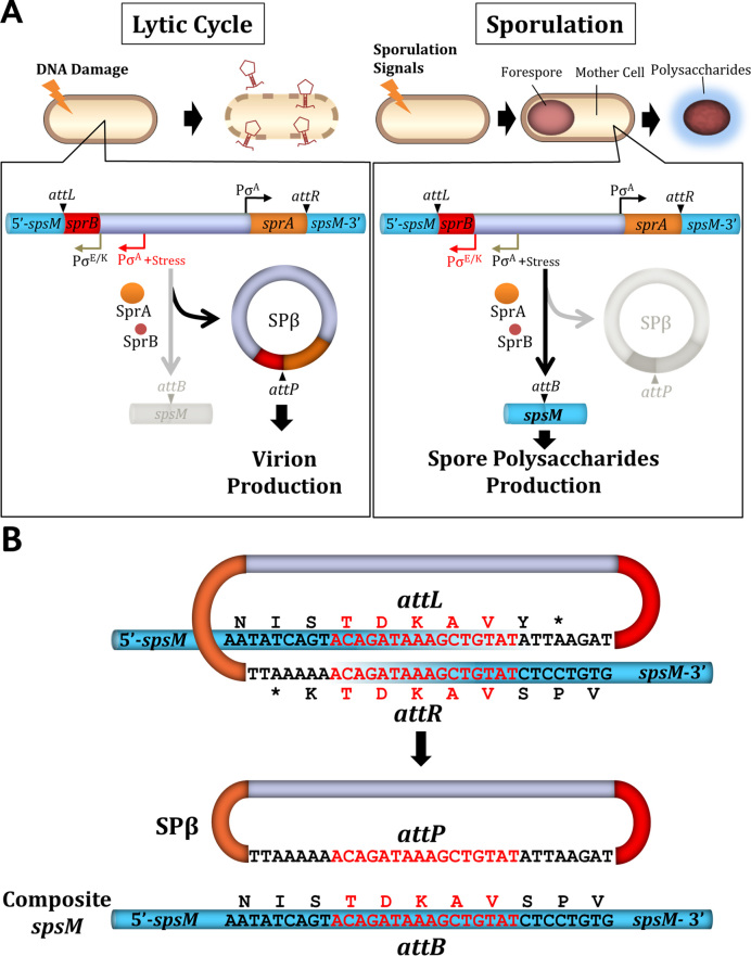
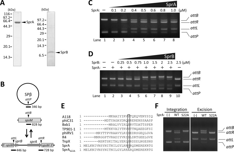
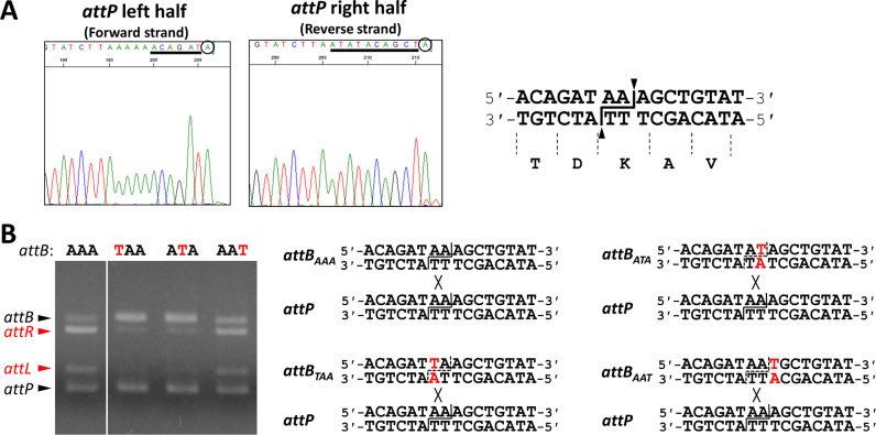
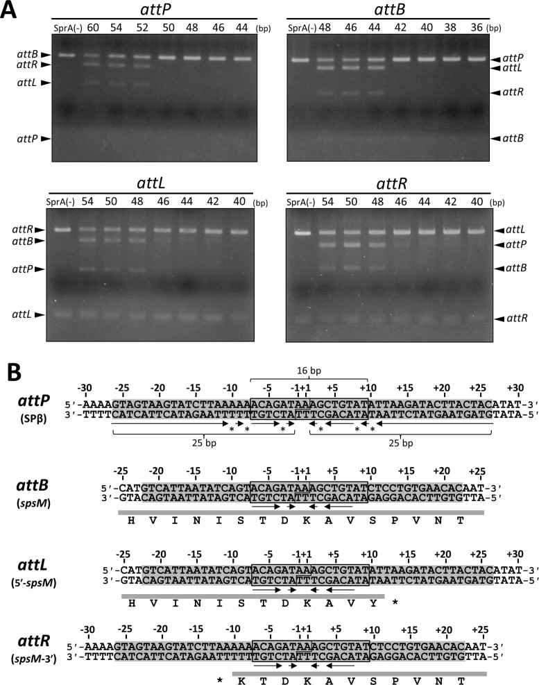
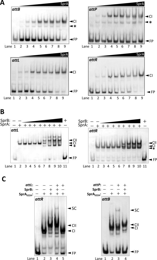
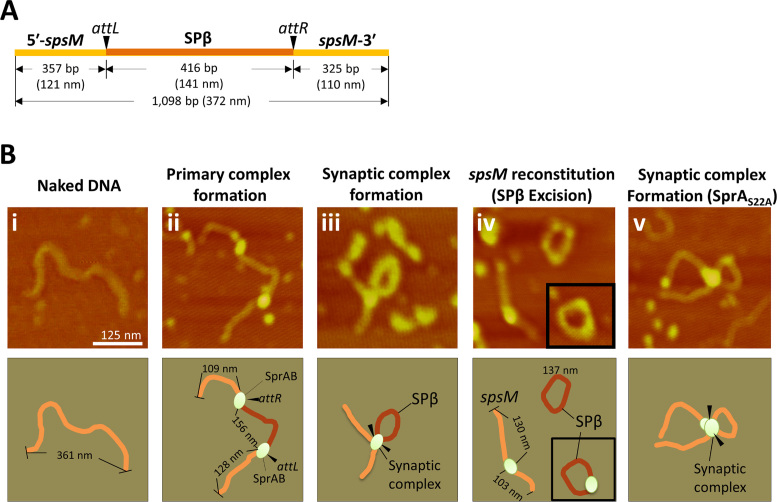
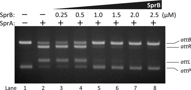
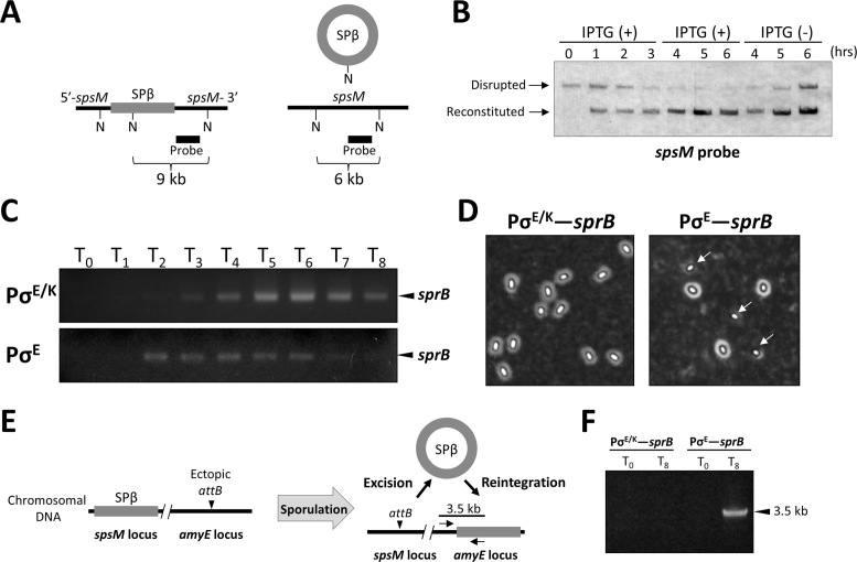
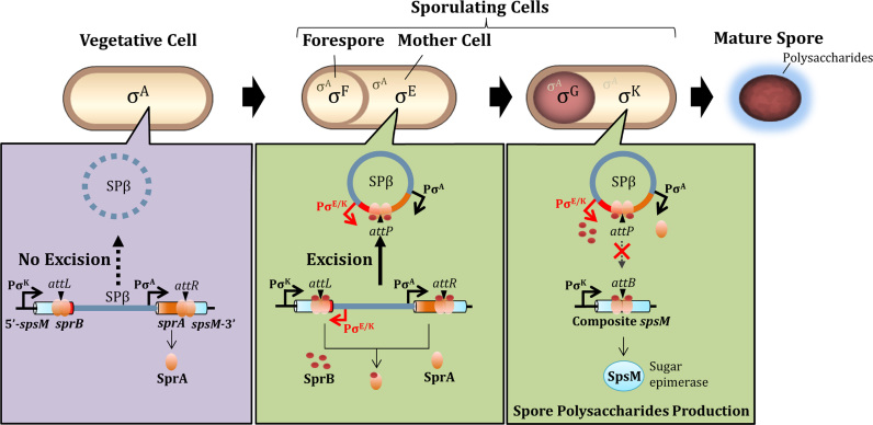
A sporulation-specific gene, spsM, is disrupted by an active prophage, SPβ, in the genome of Bacillus subtilis. SPβ excision is required for two critical steps: the onset of the phage lytic cycle and the reconstitution of the spsM-coding frame during sporulation. Our in vitro study demonstrated that SprA, a serine-type integrase, catalyzed integration and excision reactions between attP of SPβ and attB within spsM, while SprB, a recombination directionality factor, was necessary only for the excision between attL and attR in the SPβ lysogenic chromosome. DNA recombination occurred at the center of the short inverted repeat motif in the unique conserved 16 bp sequence among the att sites (5΄-ACAGATAA/AGCTGTAT-3΄; slash, breakpoint; underlines, inverted repeat), where SprA produced the 3΄-overhanging AA and TT dinucleotides for rejoining the DNA ends through base-pairing. Electrophoretic mobility shift assay showed that SprB promoted synapsis of SprA subunits bound to the two target sites during excision but impaired it during integration. In vivo data demonstrated that sprB expression that lasts until the late stage of sporulation is crucial for stable expression of reconstituted spsM without reintegration of the SPβ prophage. These results present a deeper understanding of the mechanism of the prophage-mediated bacterial gene regulatory system.


Figures

Figure 1.
Figure 1.
SPβ excision and spsM rearrangement. (A) Diagram of SPβ prophage excision in B. subtilis 168. In the lytic cycle, the excised SPβ DNA is incorporated into the phage capsids to produce the virion and the host cell undergoes lysis. During sporulation, the prophage excision generates functional spsM, which is necessary for production of the spore surface polysaccharides. (B) Nucleotide sequences of the junctions prior and posterior to DNA recombination (21). The 16 bp overlapping nucleotide sequence is indicated in red. Deduced amino-acids sequences are shown above or below the nucleotide sequences.
Figure 2.
Figure 2.
In vitro recombination assays. (A) Preparation of recombinant SprA and SprB. Recombinant SprA and SprB tagged with the six histidines at their C-termini were purified by affinity chromatography. Purified SprA (2 μg) and SprB (1 μg) were separated by SDS-PAGE (10% gel) and Tricine-SDS-PAGE (16.5% gel), respectively. (B) Schematic representation of the SPβ integration/excision reaction. Triangles point to the DNA cleavage sites during the recombination. Thick lines indicate regions corresponding to the DNA substrates for in vitro recombination. (C) Integration reaction. For attB and attP, 20 nM of each substrate were reacted with SprA (0, 0.1, 0.2, 0.4, 0.5, 0.6, 0.8 or 1.0 μM) at 37°C for 60 min. Reaction products were separated by 2% agarose gel electrophoresis. (D) Excision reaction. For attL and attR, 20 nM of each substrate were reacted with SprA (0 or 0.5 μM) and SprB (0, 0.25, 0.5, 0.75, 1.0, 1.5, 2.0 or 2.5 μM) at 37°C for 60 min. The reaction products were loaded on a 2% agarose gel. +/–, presence or absence of SprA (0.5 μM) or SprB (2.5 μM). (E) Alignment of amino-acid sequences of the NTDs of the LSRs. The nucleophilic serine residue (S) is indicated by the box. (F) Impact of a substitution of the 22nd serine with alanine. In vitro integration/excision recombination assays were performed using wild-type and mutated SprA (0.5 μM) and SprB (1.0 μM). (-), absence of SprA; WT, wild-type SprA; S22A, SprAS22A.
Figure 3.
Figure 3.
DNA cleavage and strand exchange by SprA. (A) Direct sequencing of the intermediates of the SprA-mediated integration reaction. The intermediates: the left and right half-sites of attP, were directly sequenced using a BigDye Terminator v3.1 Cycle Sequencing Kit and an ABI 3500 DNA analyzer. The 16 bp overlapping sequence is underlined. The 3΄-end nucleotides indicated by the circles are adenines added by the terminal transferase activity of Taq polymerase used for the cycle sequencing reaction. The cleavage point of attP is shown to the left (indicated by the lines and triangles). (B) Effects of point mutations of the central dinucleotides on DNA recombination. Point mutations were introduced into the AAA nucleotides at the center of the attB site (A→T). In vitro integration recombination was performed using 0.5 μM SprA and 50 ng each of the wild-type attP and the mutated attB substrates.
Figure 4.
Figure 4.
Determination of the minimal att sites. (A) Evaluation of the recombination activities of the various-sized att substrates. Each ∼50 ng of 40–60 bp DNA fragments containing the att sites with progressive deletion from both ends were reacted with 0.5 μM SprA in the presence of 50 ng of the ∼3 kb DNA fragments containing the intact partner att sequences (attP, 3,015 bp; attB, 3,190 bp; attL, 3,221 bp; attR, 2,984 bp). Nucleotide sequences of the short att substrates are shown in (B). Schematic of the recombination reaction is illustrated in Supplementary Figure S4. Numbers above the panels indicate the sizes of the short att fragments. SprA (–) indicates the absence of SprA. (B) Nucleotide sequences of the minimal att sites. Nucleotide sequences of the four att sites are shown with their positions. The positions of −1 and +1 are assigned to the central nucleotides of the attP site. The minimal att sites are indicated by shading. The 16 bp consensus sequences are indicated by boxes. Arrows and asterisks denote the inverted repeat sequences and the mismatched nucleotides within the attP sites, respectively. The deletion series of the short att substrates were shown as below: attP60, from −30 to +30; attP54, −27 to +27; attP52, −26 to +26; attP50, −25 to +25; attP48, −24 to +24; attP46, −23 to +23; attP44, −22 to +22; attB48, −24 to +24; attB46, −23 to +23; attB44, −22 to +22; attB42, −21 to +21; attB40, −20 to +20; attB38, −19 to +19; attB36, −18 to +18; attL54, −25 to +29; attL50, −23 to +27; attL48, −22 to +26; attL46, −21 to +25; attL44, −20 to +24; attL42, −19 to +23; attL40, −18 to +22; attR54, −29 to +25; attR50, −27 to +23; attR48, −26 to +22; attR46, −25 to +21; attR44, −24 to +20; attR42, −23 to +19; attR40, −22 to +18.
Figure 5.
Figure 5.
Electrophoretic mobility shift assays (A) SprA-DNA complex formation. For these assays, 10 nM of the DIG-labeled attP (94 bp), attB (111 bp), attL (116 bp), and attR (137 bp) probes were reacted with the various concentrations of SprA at 37°C for 30 min. SprA concentrations were as follows: 0, 25, 50, 100, 150, 200, 250, 300 and 350 nM. The reaction mixtures were separated by 4% native gels. FP, free probe; CI, SprA–DNA complex. Asterisks indicate SprA monomers bound to DNA. (B) SprA–SprB-DNA complex formation. The attL and attR probes (10 nM) were reacted with 0.4 μM SprA in the presence of 0.1, 0.2, 0.4, 0.8, 1.6, 3.2, 6.4 or 12.8 μM SprB (lanes 3–10). FP, free probe; CI, SprA–DNA complex; CII, SprA–SprB–DNA complex; *, additional bands of unknown nature; +/−, presence or absence of 0.4 μM SprA and 12.8 μM SprB. (C) Synaptic complex formation. Here, 10 nM of each of the attR (left panel) and attB probes (right panel) were reacted with 0.4 μM SprAS22A in the presence or absence of 3.2 μM SprB and 20 nM non-labeled partner att DNA. FP, free probe; CI, SprA-DNA complexes; CII, SprA–SprB-DNA complexes; SC, synaptic complexes; +/−, presence or absence of 0.4 μM SprAS22A, 3.2 μM SprB, and 20 nM non-labeled att DNA.
Figure 6.
Figure 6.
AFM imaging of the spsM rearrangement. (A) Schematic of a 1098-bp DNA substrate for AFM imaging. Size (nm) of the DNA molecule was calculated from one DNA base pair of 0.34 nm in length (68). (B) Representative images at each of the stages during the excision reaction. The DNA substrate (4.8 nM) was incubated with or without SprA (0.5 μM) and SprB (1.6 μM) at 37°C for 15 min and then observed using AFM. The top panels show the AFM images: i, naked DNA; ii, primary complex; iii, synaptic complex; iv, excised SPβ and reconstituted spsM; v, the synaptic complex containing SprAS22A. Scale bar indicates 125 nm. The bottom panels are illustrations of the AFM images. Actual sizes (nm) of the DNA molecules were measured from AFM images.
Figure 7.
Figure 7.
Inhibition of integration by SprB. The attP and normal attB substrates (20 nM each) were reacted with 0.5 μM SprA in the presence of the various concentrations of SprB (0, 0.25, 0.5, 1.0, 1.5, 2.0 or 2.5 μM) at 37°C for 60 min.
Figure 8.
Figure 8.
Repression of reintegration by SprB. (A) A schematic shows the B. subtilis spsM locus prior (left) and posterior (right) to the SPβ excision. Black and gray lines represent the spsM gene and the SPβ prophage region. N, NdeI-recognition sites; thick line, spsM-specific probe for Southern blotting. (B) Artificial control of SPβ excision and integration. The BsINDB strain harboring an IPTG-inducible sprB construct was cultured in the presence and absence of 0.25 mM IPTG, as described in the Materials and Methods section. Chromosomal DNA was extracted from the cells at indicated time points after the addition of IPTG (T = 0 h) and subjected to Southern hybridization. Disrupted, disrupted spsM (9 kb); Reconstituted, reconstituted spsM (6 kb). (C) Transcription of sprB during sporulation. Total RNA was isolated from the 168 and BSIID sporulating cells. The sprB cDNA was synthesized from 1 μg of total RNA, amplified by 20 cycles of PCR, and analyzed using 1.5% agarose gel electrophoresis. T0–8 denote the times after the onset of sporulation. Arrowheads indicate the RT-PCR products of sprB (D) Effect of a change in sprB expression pattern on spore morphology. Spores produced by the strain 168 (PσE/K–sprB) and BSIID (PσE–sprB) cells were negatively stained with Indian ink and observed using phase-contrast microscopy. Arrows indicate spores that lack the polysaccharide layer. (E) Schematic of the SPβ reintegration assay. B. subtilis strains, 168-AEB (PσE/K–sprB) and BSIID-AEB (PσE–sprB) possess an ectopic attB at the amyE locus. Reintegration of the excised SPβ into the amyE locus was detected by PCR using a combination of the sprB- and amyE-specific primers. (F) Detection of the reintegration. PCR was performed using chromosomal DNA from the 168-AEB and BSIID-AEB cells at T0 and T8 with the primers shown in the panel E. The arrowhead indicates the PCR product that corresponds to the 3.5 kb region within the amyE–sprB locus.
Figure 9.
Figure 9.
A model for control of the irreversible regulatory switch for spsM. The schematic representation shows morphological changes during sporulation and the spsM rearrangement in B. subtilis 168. The housekeeping (σA) and sporulation-specific (mother cell, σE→σK; forespore, σF→σG) sigma factors are indicated in the schematic. In the SPβ lysogen, sprA is constitutively expressed in the vegetative phase; nevertheless, the excisional recombination does not occur due to the lack of SprB. During sporulation, SprB is expressed from the mid until the late stages and participates in the excision of SPβ prophage. The excised SPβ is prevented from reintegrating because the SprA–SprB complex cannot catalyze the integrative recombination. The inhibitory effect of SprB on reintegration allows the stable expression of functional spsM during sporulation. Stoichiometry of SprA and SprB in the complex is not considered in this cartoon.

References

    1. Sakano H., Rogers J.H., Huppi K., Brack C., Traunecker A., Maki R., Wall R., Tonegawa S.. Domains and the hinge region of an immunoglobulin heavy chain are encoded in separate DNA segments. Nature. 1979; 277:627–633. - PubMed
    1. Schatz D.G., Ji Y.. Recombination centres and the orchestration of V(D)J recombination. Nat. Rev. Immunol. 2011; 11:251–263. - PubMed
    1. Schatz D.G., Oettinger M.A., Baltimore D.. The V(D)J recombination activating gene, RAG-1. Cell. 1989; 59:1035–1048. - PubMed
    1. Oettinger M.A., Schatz D.G., Gorka C., Baltimore D.. RAG-1 and RAG-2, adjacent genes that synergistically activate V(D)J recombination. Science. 1990; 248:1517–1523. - PubMed
    1. Alt F.W., Zhang Y., Meng F.L., Guo C., Schwer B.. Mechanisms of programmed DNA lesions and genomic instability in the immune system. Cell. 2013; 152:417–429. - PMC - PubMed