. 2017 Jul 1;23(7):461-477.
doi: 10.1093/molehr/gax029.

Intergenerational impact of paternal lifetime exposures to both folic acid deficiency and supplementation on reproductive outcomes and imprinted gene methylation

Affiliations

Intergenerational impact of paternal lifetime exposures to both folic acid deficiency and supplementation on reproductive outcomes and imprinted gene methylation

Lundi Ly et al. Mol Hum Reprod. .

Abstract

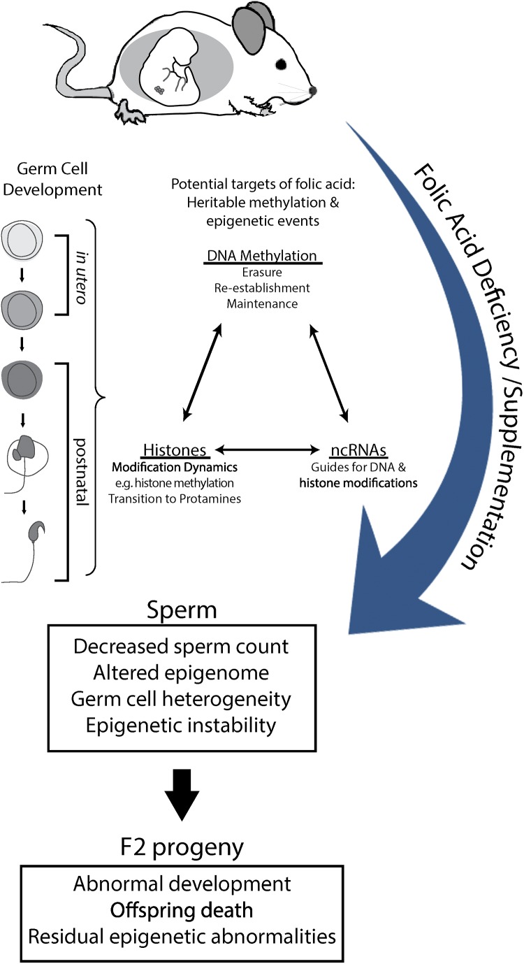
Study question: Do paternal exposures to folic acid deficient (FD), and/or folic acid supplemented (FS) diets, throughout germ cell development adversely affect male germ cells and consequently offspring health outcomes?

Summary answer: Male mice exposed over their lifetimes to both FD and FS diets showed decreased sperm counts and altered imprinted gene methylation with evidence of transmission of adverse effects to the offspring, including increased postnatal-preweaning mortality and variability in imprinted gene methylation.

What is known already: There is increasing evidence that disruptions in male germ cell epigenetic reprogramming are associated with offspring abnormalities and intergenerational disease. The fetal period is the critical time of DNA methylation pattern acquisition for developing male germ cells and an adequate supply of methyl donors is required. In addition, DNA methylation patterns continue to be remodeled during postnatal spermatogenesis. Previous studies have shown that lifetime (prenatal and postnatal) folic acid deficiency can alter the sperm epigenome and increase the incidence of fetal morphological abnormalities.

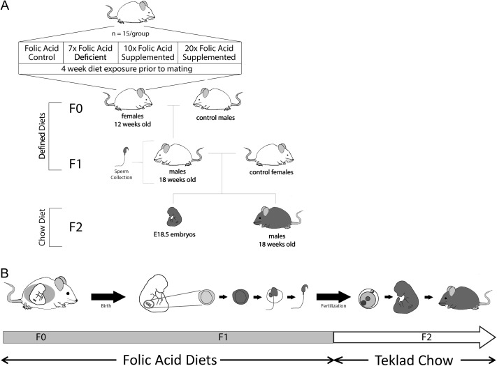
Study design, size, duration: Female BALB/c mice (F0) were placed on one of four amino-acid defined diets for 4 weeks before pregnancy and throughout pregnancy and lactation: folic acid control (Ctrl; 2 mg/kg), 7-fold folic acid deficient (7FD; 0.3 mg/kg), 10-fold high FS (10FS, 20 mg/kg) or 20-fold high FS (20FS, 40 mg/kg) diets. F1 males were weaned to their respective prenatal diets to allow for diet exposure during all windows of germline epigenetic reprogramming: the erasure, re-establishment and maintenance phases.

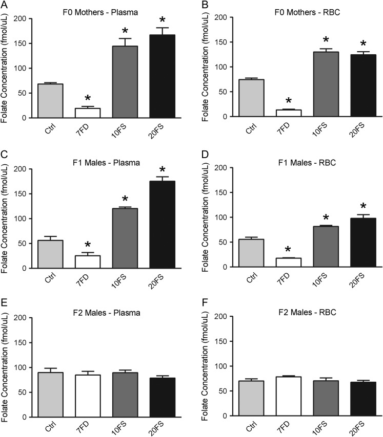
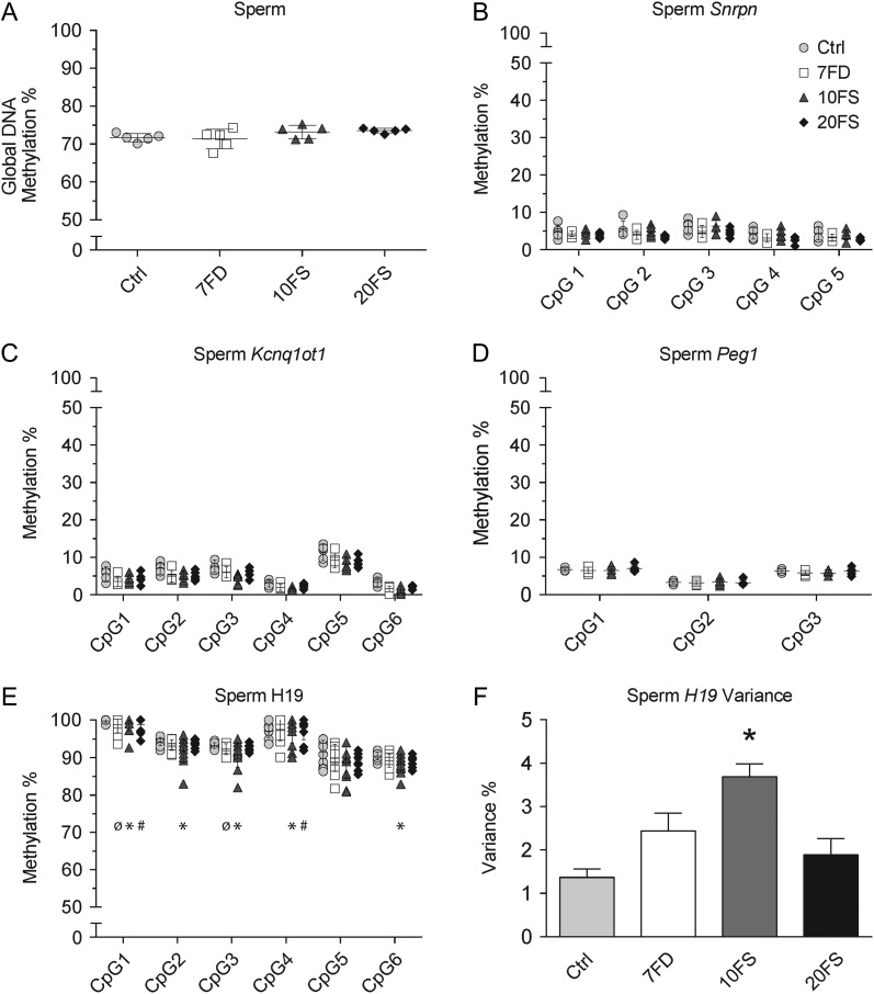
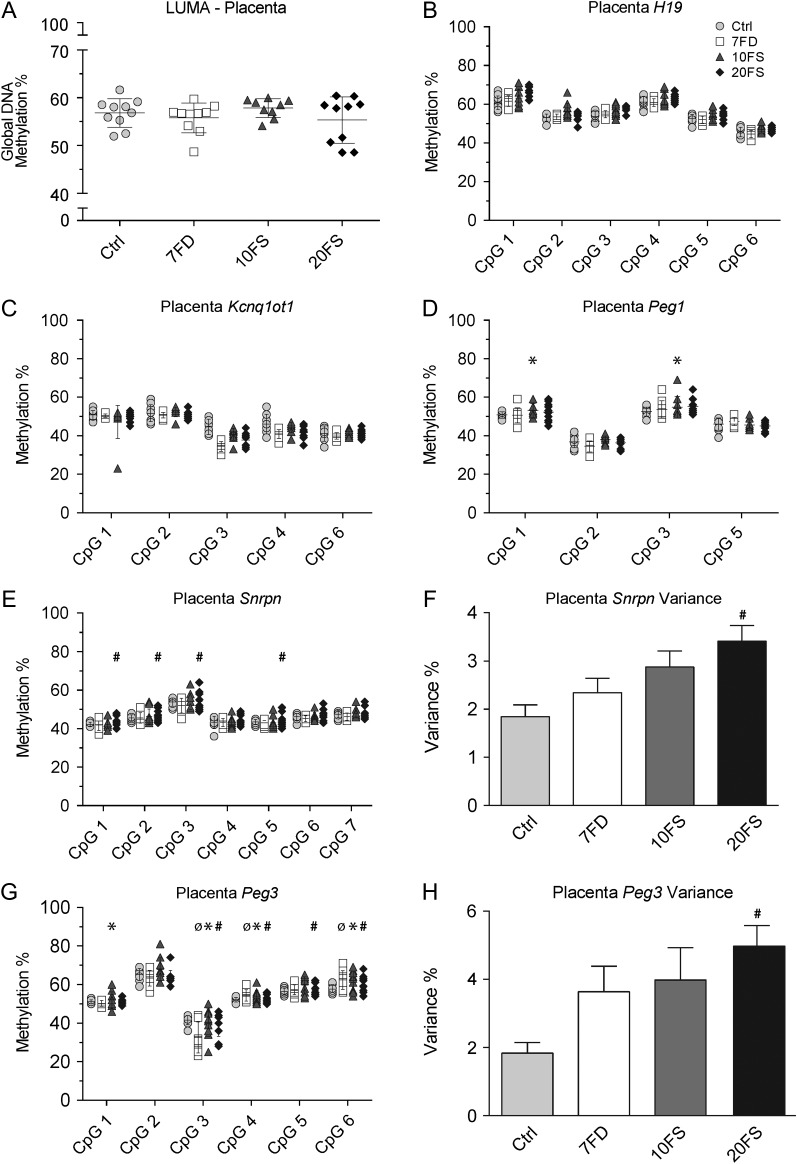
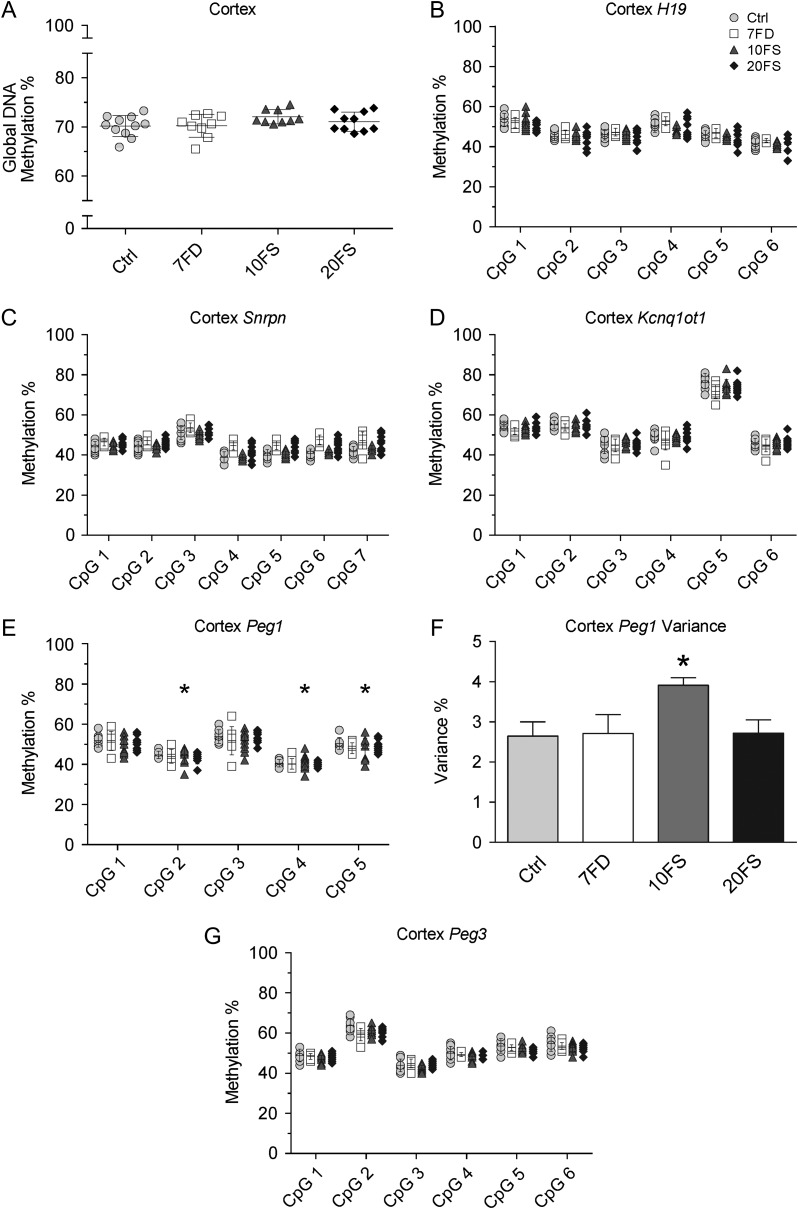
Participants/materials, settings, methods: F0 females were mated with chow-fed males to produce F1 litters whose germ cells were exposed to the diets throughout embryonic development. F1 males were subsequently mated with chow-fed female mice. Two F2 litters, unexposed to the experimental diets, were generated from each F1 male; one litter was collected at embryonic day (E)18.5 and one delivered and followed postnatally. DNA methylation at a global level and at the differentially methylated regions of imprinted genes (H19, Imprinted Maternally Expressed Transcript (Non-Protein Coding)-H19, Small Nuclear Ribonucleoprotein Polypeptide N-Snrpn, KCNQ1 Opposite Strand/Antisense Transcript 1 (Non-Protein Coding)-Kcnq1ot1, Paternally Expressed Gene 1-Peg1 and Paternally Expressed Gene 3-Peg3) was assessed by luminometric methylation analysis and bisulfite pyrosequencing, respectively, in F1 sperm, F2 E18.5 placenta and F2 E18.5 brain cortex.

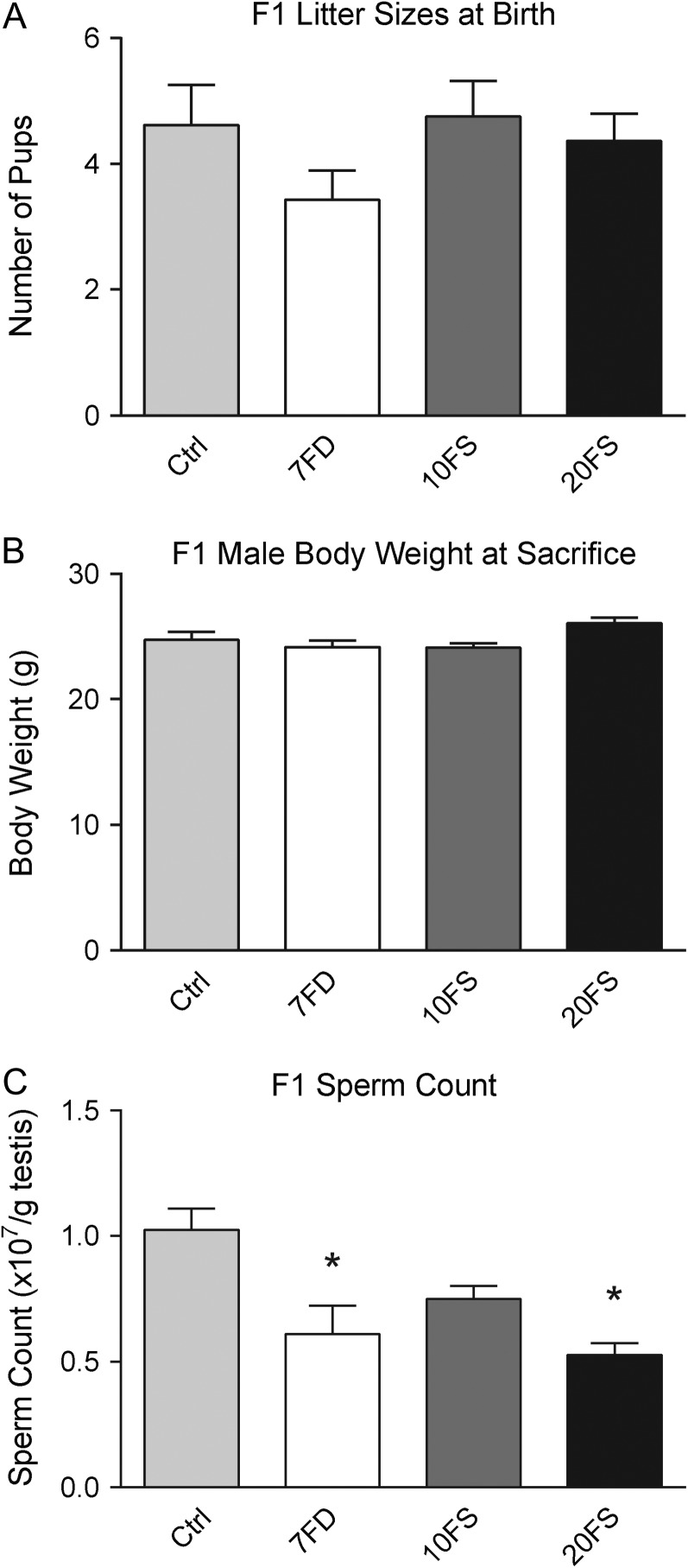
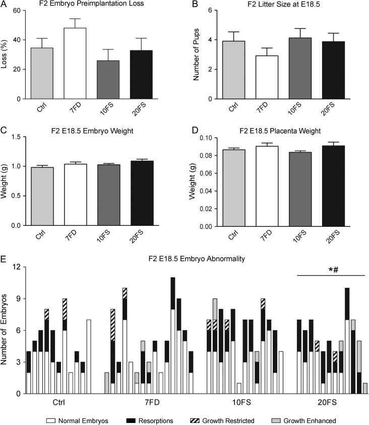
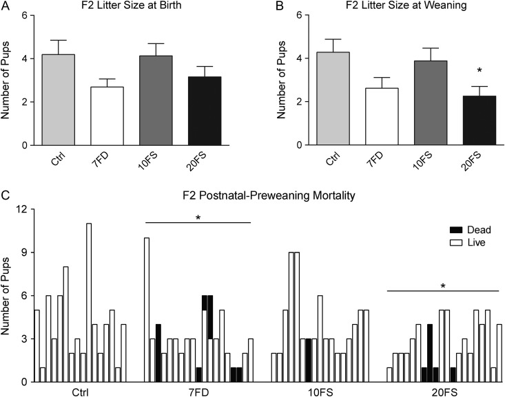
Main results and the role of chance: F1 males exhibited lower sperm counts following lifetime exposure to both folic acid deficiency and the highest dose of folic acid supplementation (20FS), (both P < 0.05). Post-implantation losses were increased amongst F2 E18.5 day litters from 20FS exposed F1 males (P < 0.05). F2 litters derived from both 7FD and 20FS exposed F1 males had significantly higher postnatal-preweaning pup death (both P < 0.05). Sperm from 10FS exposed males had increased variance in methylation across imprinted gene H19, P < 0.05; increased variance at a few sites within H19 was also found for the 7FD and 20FS groups (P < 0.05). While the 20FS diet resulted in inter-individual alterations in methylation across the imprinted genes Snrpn and Peg3 in F2 E18.5 placenta, ≥50% of individual sites tested in Peg1 and/or Peg3 were affected in the 7FD and 10FS groups. Inter-individual alterations in Peg1 methylation were found in F2 E18.5 day 10FS group brain cortex (P < 0.05).

Large scale data: Not applicable.

Limitations reasons for caution: The cause of the increase in postnatal-preweaning mortality was not investigated post-mortem. Further studies are required to understand the mechanisms underlying the adverse effects of folic acid deficiency and supplementation on developing male germ cells. Genome-wide DNA and histone methylome studies as well as gene expression studies are required to better understand the links between folic acid exposures, an altered germ cell epigenome and offspring outcomes.

Wider implications of the findings: The findings of this study provide further support for paternally transmitted environmental effects. The results indicate that both folic acid deficiency and high dose supplementation can be detrimental to germ cell development and reproductive fitness, in part by altering DNA methylation in sperm.

Study funding and competing interests: This study was supported by a grant to J.M.T. from the Canadian Institutes of Health Research (CIHR #89944). The authors declare they have no conflicts of interest.

Keywords: DNA methylation; developmental programming; epigenetics; folate; folic acid; intergenerational effects; male-mediated; paternal effects; sperm.


Figures

Figure 1
Figure 1
Intergenerational reproductive effects of lifetime folic acid deficiency and supplementation. A) Mating scheme for intergenerational exposures to diet. Eight-week-old BALB/c F0 females were fed either a Ctrl, 7FD, 10FS or 20FS diet, (n = 15 for each) for 4 weeks prior to breeding with BALB/c males fed with regular rodent chow. Females were killed, and F1 male pups received the same experimental diet as their mother. At 18 weeks of age, one F1 male from each litter was mated with a female fed with rodent chow. Females were maintained on the rodent chow through pregnancy and lactation. From weaning until death, F2 male pups received rodent chow. B) Time line of exposure to folic acid defined diets. (Ctrl, folic acid control diet; 7FD, 7× folic acid deficient; 10FS, 10× FS; 20FS, 20× FS). FS, folic acid supplemented; FD, folate deficient.
Figure 2
Figure 2
Plasma and RBC folate concentrations. Plasma and RBC folate concentrations in F0 dams (AB), F1 male progeny (CD) and F2 male progeny (EF) when killed (n = 5). *P < 0.05 by one-way ANOVA with Dunnett's multiple comparisons test versus Ctrl. RBC, red blood cell.
Figure 3
Figure 3
Lifetime folic acid deficiency and 20FS supplementation decrease sperm count. Effect of lifetime folic acid deficiency and supplementation on F1 litter sizes at birth (A; n = 11–15 litters/group), F1 adult male body weight (B; n = 17–20/group) and F1 sperm count (C; n = 6/group). *P < 0.05 by one-way ANOVA with Dunnett's multiple comparisons test.
Figure 4
Figure 4
Effects of lifetime folic acid deficiency and supplementation on pregnancy outcomes at E18.5. Preimplantation loss of F2 at embryonic day (E)18.5 (A; n = 11–15 F2 litters, representing n = 7–9 original F1 litters), F2 litter sizes at E18.5 (B), F2 embryo weights at day 18.5 (C; n = 25–38 embryos, representing n = 7–9 original F1 litters), and F2 placental weights at embryonic day 18.5 (D). Incidence of fetal abnormalities at E18.5 per litter (E; n = 11–15 litters); growth restriction and enhancement are defined as a 2-fold SD difference of embryo weight to the group mean of litter mean weights. *P < 0.05 by one-way ANOVA with Dunnett's multiple comparisons test.
Figure 5
Figure 5
Lifetime folic acid deficiency and 20FS supplementation result in postnatal-preweaning mortality. Effect of lifetime folic acid deficiency and supplementation on F2 litter sizes at birth (A; n = 16–20 F2 litters, representing n = 9–11 original F1 litters), F2 litter sizes at weaning (PND21) (B), and incidence of pup postnatal mortality per litter (C). *P < 0.05 by one-way ANOVA with Dunnett's multiple comparisons test or by Fisher's exact test.
Figure 6
Figure 6
F1 sperm global DNA methylation and DMR methylation at imprinted genes uncovers variance of H19 methylation. Global DNA methylation was measured by LUMA (A). Imprinted gene methylation was quantified using bisulfite pyrosequencing at maternally methylated genes Snrpn (B; n = 5/group), Kcnq1ot1 (C; n = 5/group) and Peg1 (D; n = 5/group) and paternally methylated gene H19 (E; n = 10–13/group). H19 variance (F) was measured as a mean of variances of all six CpGs. For individual CpG variance (panel E), ø (7FD), * (10FS), # (20FS) = P < 0.05 by F-test between deficient or supplemented versus control diet groups. For across-locus variance analysis (panel F), *P < 0.05 by one-way ANOVA with Dunnett's multiple comparisons test. LUMA, luminometric methylation analysis; DMR, differentially methylated region.
Figure 7
Figure 7
F2 E18.5 placenta global DNA methylation and DMR methylation at imprinted genes uncovers variance of Snrpn and Peg3 methylation in F2 pups sired by 20FS exposed males. Global DNA methylation was measured using LUMA (A; n = 10/group). Imprinted gene methylation was quantified using bisulfite pyrosequencing at paternally methylated gene H19 (B) and maternally methylated genes Kcnq1ot1 (C), Peg1 (D), Snrpn (E) and Peg3 (G), (n = 5/group). Snrpn and Peg3 variances were measured as a mean of variances of all seven and six CpGs, respectively. For individual CpG variance (panels D, E and G), ø (7FD), * (10FS), # (20FS) = P < 0.05 by F-test between deficient or supplemented versus control diet groups. For across-locus variance analysis (panels F and H) *P < 0.05 by one-way ANOVA with Dunnett's multiple comparisons test.
Figure 8
Figure 8
F2 E18.5 cortex global DNA methylation and DMR methylation at imprinted genes uncovers variance of Peg1 methylation in F2 pups sired by 10FS exposed males. Global DNA methylation was measured using LUMA (A, n = 10/group). Loci of paternally methylated gene H19 (B) and maternally methylated genes Snrpn (C), Kcnq1ot1 (D), Peg1 (E) and Peg3 (G) methylation levels were quantified by pyrosequencing (n = 5/group). Peg1 variance was measured as a mean of variances of all five CpGs. For individual CpG variance (panel E), * (10FS) = P < 0.05 by F-test between deficient or supplemented versus control diet groups. For across-locus variance analysis (panels F) *P < 0.05 by one-way ANOVA with Dunnett's multiple comparisons test.
Figure 9
Figure 9
Model of postulated epigenetic mechanisms and interactions underlying the adverse outcomes in offspring of males exposed to FD and FS diets, starting from fertilization. Lifetime exposure to both FD and FS spans two vulnerable windows of epigenetic reprogramming during germ cell development: in utero and postnatal. Here, in each window of development, both window-specific (e.g. DNA methylation erasure and histone-protamine transition) and development-long (e.g. DNA methylation re-establishment/maintenance and histone modification dynamics) events are at risk of being influenced by external exposures of folic acid. Interactions between DNA methylation, histone modifications and ncRNA expression all have the potential to contribute to the decreased reproductive fitness of sperm, observed as decreased sperm count, altered epigenome (DNA methylation variance), germ cell heterogeneity and epigenetic instability. Ultimately, the cumulative and interacting epigenetic effects of lifetime exposures on the sperm manifest in the F2 progeny as abnormal development, increased offspring death and residual epigenetic abnormalities. non-coding RNA, ncRNA.

Similar articles

Cited by

References

    1. Aarabi M, San Gabriel MC, Chan D, Behan NA, Caron M, Pastinen T, Bourque G, MacFarlane AJ, Zini A, Trasler J. High-dose folic acid supplementation alters the human sperm methylome and is influenced by the MTHFR C677T polymorphism. Hum Mol Genet 2015;24:6301–6313. - PMC - PubMed
    1. Bailey RL, Mills JL, Yetley EA, Gahche JJ, Pfeiffer CM, Dwyer JT, Dodd KW, Sempos CT, Betz JM, Picciano MF. Unmetabolized serum folic acid and its relation to folic acid intake from diet and supplements in a nationally representative sample of adults aged > or =60 y in the United States. Am J Clin Nutr 2010;92:383–389. - PMC - PubMed
    1. Bailey SW, Ayling JE. The extremely slow and variable activity of dihydrofolate reductase in human liver and its implications for high folic acid intake. Proc Natl Acad Sci USA 2009;106:15424–15429. - PMC - PubMed
    1. Barua S, Kuizon S, Brown WT, Junaid MA. DNA methylation profiling at single-base resolution reveals gestational folic acid supplementation influences the epigenome of mouse offspring cerebellum. Front Neurosci 2016;10:168. - PMC - PubMed
    1. Barua S, Kuizon S, Chadman KK, Flory MJ, Brown WT, Junaid MA. Single-base resolution of mouse offspring brain methylome reveals epigenome modifications caused by gestational folic acid. Epigenetics Chromatin 2014;7:3. - PMC - PubMed

Publication types

MeSH terms

Grants and funding