. 2017 Jul 7;429(14):2192-2210.
doi: 10.1016/j.jmb.2017.05.015. Epub 2017 May 20.

Structural Basis for Asymmetric Conductance of the Influenza M2 Proton Channel Investigated by Solid-State NMR Spectroscopy

Affiliations

Structural Basis for Asymmetric Conductance of the Influenza M2 Proton Channel Investigated by Solid-State NMR Spectroscopy

Venkata S Mandala et al. J Mol Biol. .

Abstract

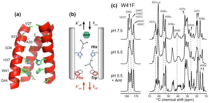
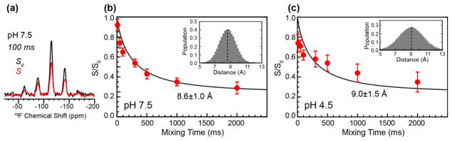
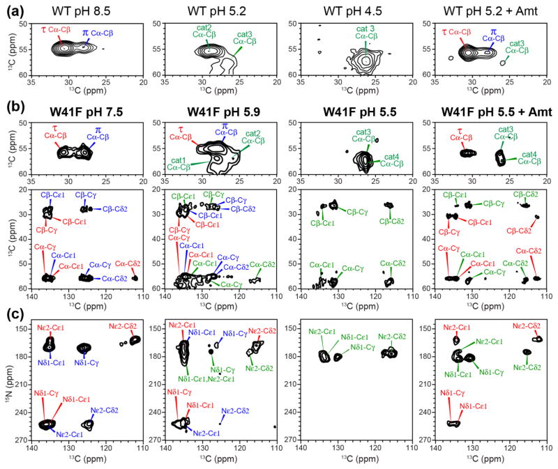
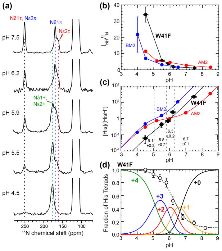
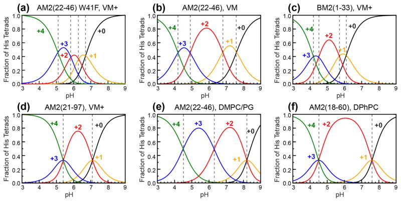
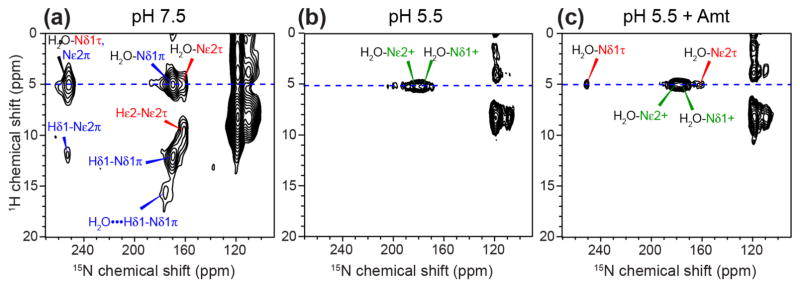
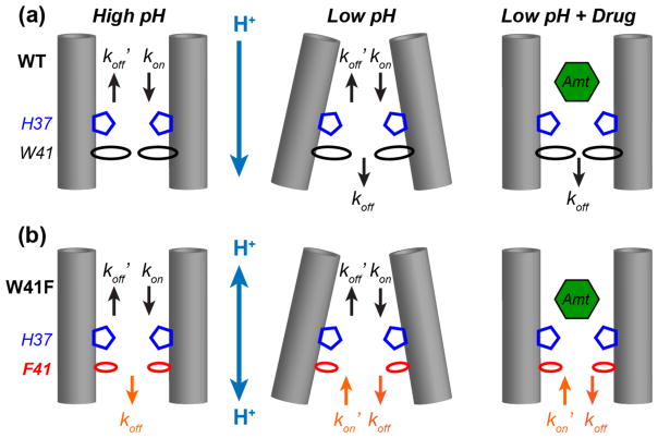
The influenza M2 protein forms an acid-activated proton channel that is essential for virus replication. The transmembrane H37 selects for protons under low external pH while W41 ensures proton conduction only from the N terminus to the C terminus and prevents reverse current under low internal pH. Here, we address the molecular basis for this asymmetric conduction by investigating the structure and dynamics of a mutant channel, W41F, which permits reverse current under low internal pH. Solid-state NMR experiments show that W41F M2 retains the pH-dependent α-helical conformations and tetrameric structure of the wild-type (WT) channel but has significantly altered protonation and tautomeric equilibria at H37. At high pH, the H37 structure is shifted toward the π tautomer and less cationic tetrads, consistent with faster forward deprotonation to the C terminus. At low pH, the mutant channel contains more cationic tetrads than the WT channel, consistent with faster reverse protonation from the C terminus. 15N NMR spectra allow the extraction of four H37 pKas and show that the pKas are more clustered in the mutant channel compared to WT M2. Moreover, binding of the antiviral drug, amantadine, at the N-terminal pore at low pH did not convert all histidines to the neutral state, as seen in WT M2, but left half of all histidines cationic, unambiguously demonstrating C-terminal protonation of H37 in the mutant. These results indicate that asymmetric conduction in WT M2 is due to W41 inhibition of C-terminal acid activation by H37. When Trp is replaced by Phe, protons can be transferred to H37 bidirectionally with distinct rate constants.

Keywords: gating; ion channels; magic-angle-spinning NMR; proton dissociation equilibrium; tautomeric equilibrium.


Figures

Figure 1
Figure 1
(a) Wild-type AM2-TM peptide structure (PDB: 3LBW) showing the locations of key residues examined in this study. One of the four chains is omitted for clarity. (b) Schematic of the channel topology, showing four possible rate constants for H37-mediated proton conduction: forward protonation (kon), forward deprotonation (koff), reverse protonation ( kon), and reverse deprotonation ( koff). Mutation of W41 changes the magnitude of these rate constants. (c) 13C MAS spectra of W41F AM2-TM bound to the VM+ membrane at pH 7.5, pH 5.5, and pH 5.5 with bound amantadine (Amt). The spectra were measured at 273 K.
Figure 2
Figure 2
Aliphatic regions of 2D 13C-13C (top) and 15N-13C (bottom) correlation spectra of W41F AM2-TM at (a) pH 7.5, (b) pH 5.5, and (c) pH 5.5 with bound Amt. Assignments for H37 cross peaks are shown in red, blue and green for the τ tautomer, π tautomer, and cationic states, respectively.
Figure 3
Figure 3
19F CODEX data of 4-19F-Phe41 in W41F AM2-TM at pH 7.5 and pH 4.5. (a) Representative 19F CODEX S0 and S spectra at a mixing time of 100 ms for the pH 7.5 sample. CODEX intensities as a function of mixing time are shown for the pH 7.5 sample (b) and for the pH 4.5 sample (c). Best fits used a Gaussian distribution of distances (inset). The mean and standard deviation of the distributions are 8.6 Å and 1.0 Å for (b) and 9.0 Å and 1.5 Å for (c).
Figure 4
Figure 4
H37 chemical shifts in WT (a) and W41F (b, c) AM2-TM from 2D 13C-13C (a, b) and 15N-13C (c) correlation spectra as a function of pH. (a) H37 Cα-Cβ regions of the 2D 13C-13C spectra of the WT peptide. (b) H37 Cα-Cβ and aliphatic-aromatic regions of the 2D 13C-13C spectra of the W41F mutant. (c) Aromatic region of the 2D 15N-13C correlation spectra of the W41F mutant. The pH and drug binding state of the samples are indicated. The mutant channel shows higher π tautomer intensities at high pH and more cationic histidine peaks at low pH compared to the WT channel.
Figure 5
Figure 5
Quantification of H37 pKa’s in membrane-bound W41F AM2-TM by 15N NMR. (a) pH-dependent 15N spectra of H37 sidechains measured at 243–248 K. Assignment of protonated and unprotonated 15N peaks is based on 2D correlation spectra. (b) Integrated intensity ratios of protonated and unprotonated imidazole nitrogens as a function of pH. W41F AM2-TM (black) shows higher protonated nitrogen intensities at low pH compared to previously measured WT AM2-TM (red) [43] and BM2-TM results (blue) [69]. (c) Neutral-to-cationic histidine concentration ratios as a function of pH. Best fit of the data yield four pKa’s for W41F AM2-TM, which are indicated by dashed vertical lines. (d) Population distributions of charged tetrads of W41F AM2-TM. The +2 charged channel has the lowest population among the five charge states. Normalized proton conductance of W41F M2 from the literature [7] (open circles) is fit using the charge-state distribution to estimate the relative conductance of the different charge states.
Figure 6
Figure 6
Comparison of pH-dependent populations of the five charge states of various M2 samples studied so far. (a) W41F AM2(22-46) in the VM+ membrane from this study. (b) WT AM2(22-46) in the VM membrane [43]. (c) BM2(1-33) in the VM+ membrane [69]. (d) AM2(21-97) in the VM+ membrane [31]. (e) AM2(22-46) in the DMPC/DMPG membrane [47]. (f) AM2(18-60) in the DPhPC membrane [48]. Dashed vertical lines indicate the intersection of two adjacent population curves, which correspond to the pKa’s.
Figure 7
Figure 7
Hydration of the W41F AM2-TM channel probed by 2D 1H-13C correlation spectra. (a) Cα S/S0 values of W41F M2. (b) Cα S/S0 values of WT M2. (c) Cβ S/S0 values of W41F M2. (d) Cβ S/S0 values of WT M2. Green squares, red diamonds, and black circles represent low pH, drug-bound low pH, and high pH data, respectively. Open symbols indicate values with greater uncertainty. Dashed green and black lines indicate the mean S/S0 value for low and high pH, respectively. The W41F mutant has lower average hydration than the WT channel at high pH and similar average hydration as the WT at low pH. However, the C-terminus of the WT peptide is less hydrated than the N-terminus at low pH, while the W41F mutant shows similar hydration of the two termini at low pH.
Figure 8
Figure 8
2D 1H-15N correlation spectra of H37 in W41F AM2-TM at (a) pH 7.5, (b) pH 5.5, and (c) pH 5.5 with bound Amt. Water 1H cross peaks at 5.0 ppm with H37 imidazole nitrogens are detected in all three samples. A 16-ppm 1H chemical shift in the pH 7.5 sample can be assigned to the π tautomer with a strong hydrogen bond to water. The absence of HN cross peaks at pH 5.5 indicates rapid exchange of H37 with water at 263 K. Drug binding retains only water-H37 cross peaks, indicating fast exchange with C-terminal protons even when the N-terminus is blocked by drug.
Figure 9
Figure 9
Schematic models of H37 protonation and deprotonation rate constants in WT (a) and W41F (b) M2 channels. Left: high pH; Middle: low pH; Right: low-pH with bound drug. For each scenario, the protonation equilibrium of the W41F mutant channel has an additional rate constant (orange) at the C-terminus compared to the WT channel. This suppresses the high pKa’s and increases the low pKa’s in the mutant compared to the WT channel, causing more clustered protonation events in the mutant.

Similar articles

Cited by

References

    1. Hille B. Ionic channels of excitable membranes. 2. Sundeland: Sinauer Associates Inc; 1992.
    1. Pinto LH, Lamb RA. The M2 proton channels of influenza A and B viruses. J Biol Chem. 2006;281:8997–9000. - PubMed
    1. Cady SD, Luo WB, Hu FH, Hong M. Structure and Function of the Influenza A M2 Proton Channel. Biochemistry. 2009;48:7356–7364. - PMC - PubMed
    1. Hong M, DeGrado WF. Structural basis for proton conduction and inhibition by the influenza M2 protein. Protein Sci. 2012;21:1620–1633. - PMC - PubMed
    1. Wang C, Lamb RA, Pinto LH. Activation of the M(2) Ion-Channel of Influenza-Virus - a Role for the Transmembrane Domain Histidine Residue. Biophys J. 1995;69:1363–1371. - PMC - PubMed

Publication types

MeSH terms