Review
. 2017 Oct:139:161-167.
doi: 10.1016/j.visres.2017.04.011. Epub 2017 Jun 9.

Diabetic choroidopathy

Affiliations
Review

Diabetic choroidopathy

Gerard A Lutty. Vision Res. 2017 Oct.

Abstract

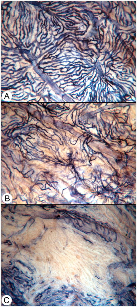
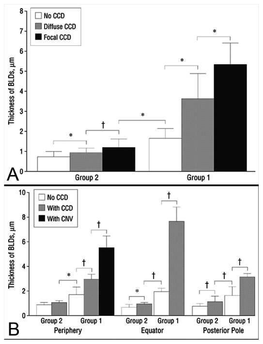
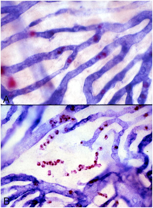
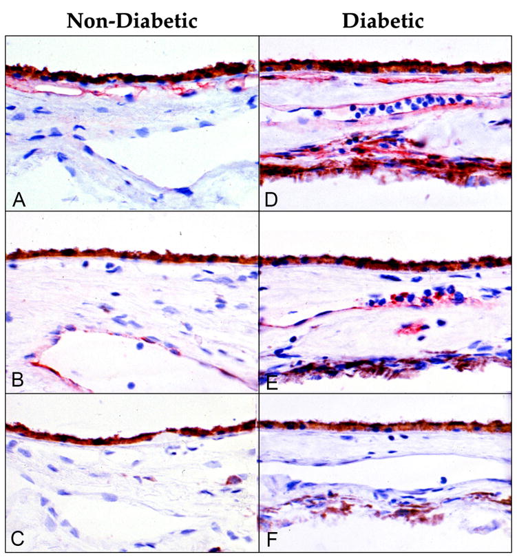
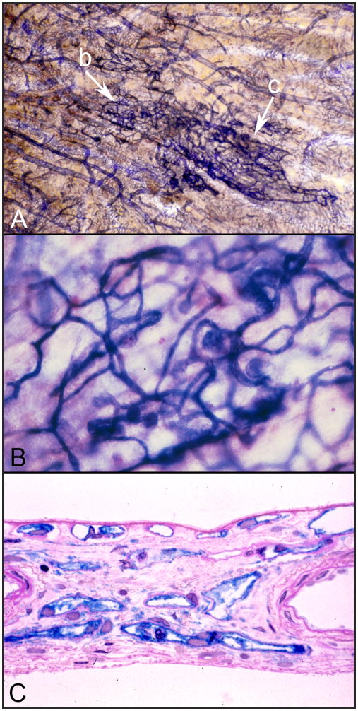
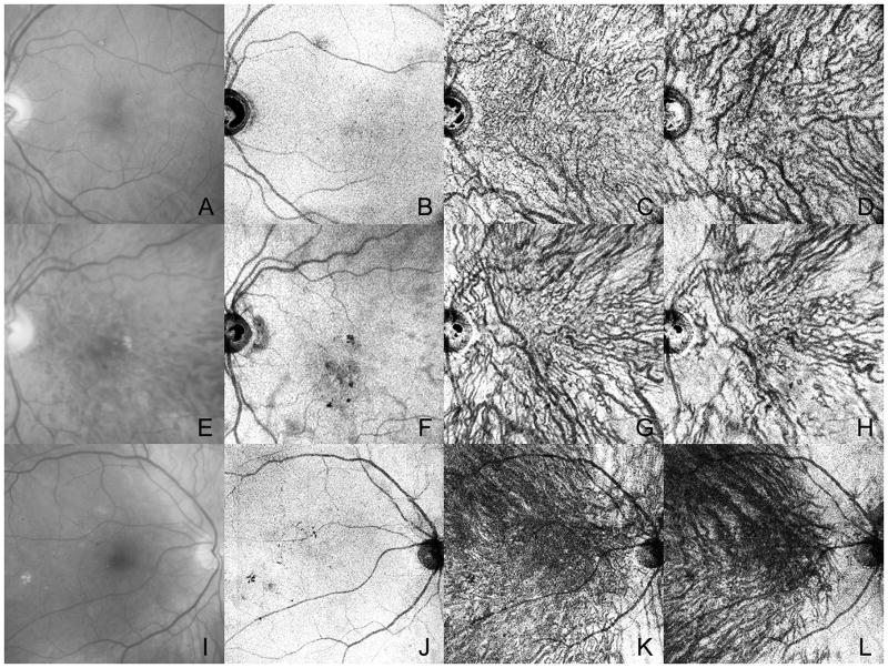
Early histopathological studies of diabetic choroids demonstrated loss of choriocapillaris (CC), tortuous blood vessels, microaneurysms, drusenoid deposits on Bruchs membrane, and choroidal neovascularization. The preponderance of histopathological changes were at and beyond equator. Studies from my lab suggest that diabetic choroidopathy is an inflammatory disease in that leukocyte adhesion molecules are elevated in the choroidal vasculature and polymorphonuclear neutrophils are often associated with sites of vascular loss. Modern imaging techniques demonstrate that blood flow is reduced in subfoveal choroidal vasculature. Angiography has shown areas of hypofluorescence and late filling that probably represent areas of vascular loss and/or compromise. Perhaps, as a result of vascular insufficiency, the choroid appears to thin in DC unless macular edema is present. Enhanced depth imaging (EDI-SD) OCT and swept source (SS) OCT have documented the tortuosity and loss in intermediate and large blood vessels in Sattler's and Haller's layer seen previously with histological techniques. The risk factors for DC include diabetic retinopathy, degree of diabetic control, and the treatment regimen. In the future, OCT angiography could be used to document loss of CC. Because most of the measurement and imaging are in the posterior pole, the severity of DC may be underappreciated in the published accounts of DC assessed with imaging techniques. However, it is now possible to document DC and quantify these changes clinically. This suggests that DC should be evaluated in future clinical trials of drugs targeting DR because vascular changes similar to those in DR are occurring in DC.

Keywords: Choriocapillaris; Choroidopathy; Diabetes mellitus; OCT; Polymorphonuclear neutrophils.


Figures

Figure 1
Figure 1
Alkaline phosphatase-incubated choroid from a diabetic choroid. (A) A normal area of choroid where all choriocapillaris are viable, i.e. are APase+. Organized lobules are apparent in this area. (B) An area in the same choroid that has diffuse choriocapillaris loss. (C) An area of complete loss of choriocapillaris. (With permission Figure 2 in Lutty, Invest. Ophthalmol. Vis. Sci. 2013; 54: ORSF81–7. doi: 10.1167/iovs.13-12979)
Figure 2
Figure 2
Choroidal neovascularization in diabetic choroid. (A) Alkaline phosphatase incubated choroid from an 74 year old type 2 diabetic that has a large CNV formation (arrow). The yellow material is basal laminar deposit, which accumulates non-reduced formazan reaction product, thus the yellow color. The CNV formation is surrounded by areas with severe CC loss. (B) A section through the CNV that was stained with PAS and hematoxylin demonstrates a pink basal laminar deposit over the CNV (arrow) which has grown through a break in Bruchs membrane (Arrowhead).
Figure 3
Figure 3
Thickness of Bruchs membrane basal laminar-like deposits (BLD) in diabetic subjects. The thickness of Bruchs membrane deposits (+/−SD) was assessed in subjects with no choriocapillaris degeneration or dropout (CCD) (white), CCD (Gray), and CCD and/or CNV (black). (A) In group 2, nondiabetic aged control subjects with no measureable CCD, there was very little deposit. In group 1, the diabetic subjects had deposits that increased in thickness as areas with CCD became more severely affected, i.e. diffuse vs complete. (B) BLDs were significantly thicker in CCD areas in periphery, equator, and posterior pole and the thickest areas had CNV. Note only periphery had CNV.(asterisk = 0.05; dagger = , 0.01)
Figure 4
Figure 4
Alkaline phosphatase/nonspecific esterase (APase/NSE) double-labeled flat mounted choroids from nondiabetic subject (A) and an insulin-dependent diabetic subject (B). APase activity (blue reaction product) is localized to viable choroidal blood vessels while NSE activity (red reaction product) labels polymorphonuclear neutrophils (PMNs) within choriocapillaris lumens and mast cells, which are larger and out of focus since they found at the levels of Sattler’s and Haller’s layers. Queues of PMNs are present in degenerating segments of capillaries in the diabetic choroid (B). Note that PMNs are present within the choriocapillaris where APase activity ceases.
Figure 5
Figure 5
Immuohistochemical localization of ICAM-1 (A, D) and P-selectin (B, E) in a nondiabetic control subject (left) and insulin dependent diabetic subject (right). (A) ICAM-1 is constitutively made by normal choriocapillaris. (B) P-selectin is localized (red reaction product) only to large veins in control subjects. (C) There is no red reaction product when non-immune IgG is used as the primary antibody. (D) ICAM-1 is elevated in CC and present in all choroid blood vessels in the diabetic subject. (E) P-selectin is present in large and intermediate blood vessels in the diabetic subject. Bright red cells in lumens of blood vessels deep in choroid are platelets. (F) There is no red reaction product in sections of the diabetic incubated with non-immune IgG. The brown cells deep in choroid are melanocytes. (Red AEC reaction product and hematoxylin counter stain) (With permission, Figure 1 from McLeod et al. Am. J. Pathol. 1995; 147:642–653)
Figure 6
Figure 6
Intrachoroidal neovascularization or microvascular abnormality (ICMA) in the choroid of a 68 year old insulin dependent diabetic. (A) The ICAM formation is the dark blue cobweb like vasculature indicated with arrows. Note that the ICMA blood vessels have the most intense APase activity. (B) Higher magnification of the portion of the ICAM formation shown at arrow “b” in the APase flat mount shown in panel “A”. This area of the formation has bulbous microaneurysms. (C) A section through the dense portion of the ICMA formation indicated by arrow “c” in panel “A”. Notice that these ICMA capillaries are intensely blue and are located in outer choroid at the level of Haller’s layer where there are normally no capillaries. (section in (C) stained with PAS and hematoxylin in addition to APase reaction product.)
Figure 7
Figure 7
Imaging of three patients with non-proliferative diabetic retinopathy. Red free images (A, E, I) of the subjects have very early changes like lipid and retinal hemorrhage. En face images from a swept source OCT is shown at different levels in the retina and choroid. (B–D) At the level of the RPE there are topographical changes due to loss of RPE (F) or shadowing from changes in sensory retina. At the level of inner choroid (C,G, K), the choriocapillaris is not clearly visible but irregular, tortuous, and beaded vessels can be seen. In outer choroid (D, H, L) enface images show attenuation of some large and intermediate vessels, especially in the subject shown in (H).(With permission, Figure 12 in Ferrara et al. Progress in Retinal and Eye Research 2016: 52:130–155)

References

    1. Adamis AP. Is diabetic retinopathy an inflammatory disease? Br J Ophthalmol. 2002;86:363–365. - PMC - PubMed
    1. Baranao RI, Garberi JC, Tesone PA, Rumi RS. Evaluation of neutrophil activity and circulating immune complexes levels in diabetic patients. Horm Metab Res. 1987;19:371–374. - PubMed
    1. Barouch F, Miyamoto K, Allport J, Fujita K, Bursell S, LP A, Luscinskas F, Adamis A. Integrin-mediated neutrophil adhesion and retinal leukostasis in diabetes. Invest Ophthalmol Vis Sci. 2000;41:1153–1158. - PubMed
    1. Campos A, Campos EJ, Martins J, Ambrosio AF, Silva R. Viewing the choroid: where we stand, challenges and contradictions in diabetic retinopathy and diabetic macular oedema. Acta Ophthalmol 2016 - PubMed
    1. Cao J, McLeod S, Merges CA, Lutty GA. Choriocapillaris degeneration and related pathologic changes in human diabetic eyes. Arch Ophthalmol. 1998;116:589–597. - PubMed

MeSH terms