. 2017 May 1;31(9):939-952.
doi: 10.1101/gad.297051.117. Epub 2017 May 23.

The LOTUS domain is a conserved DEAD-box RNA helicase regulator essential for the recruitment of Vasa to the germ plasm and nuage

Affiliations

The LOTUS domain is a conserved DEAD-box RNA helicase regulator essential for the recruitment of Vasa to the germ plasm and nuage

Mandy Jeske et al. Genes Dev. .

Abstract

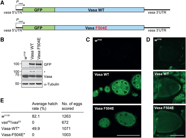
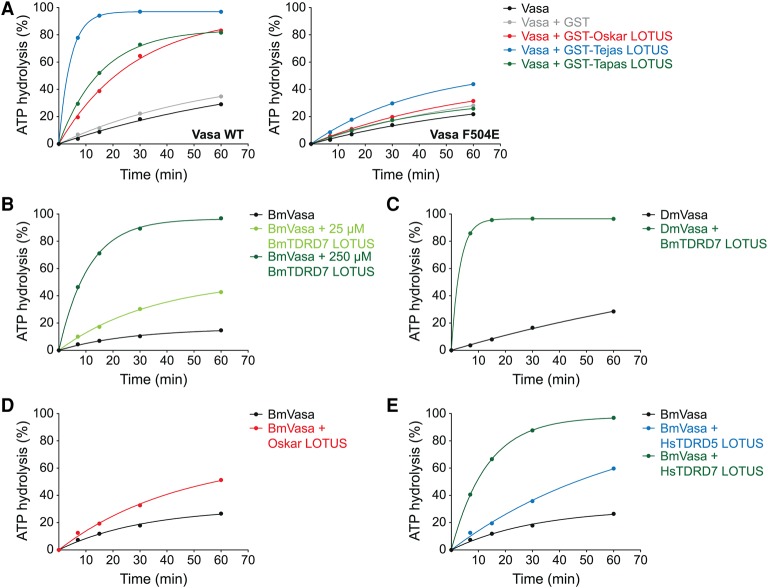
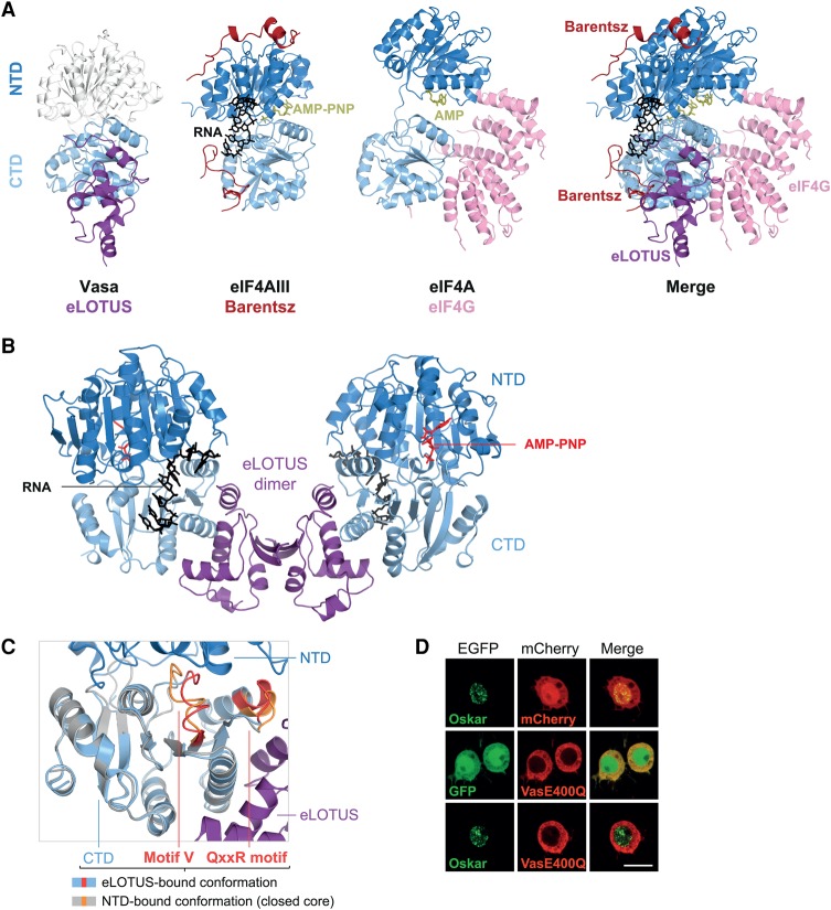
DEAD-box RNA helicases play important roles in a wide range of metabolic processes. Regulatory proteins can stimulate or block the activity of DEAD-box helicases. Here, we show that LOTUS (Limkain, Oskar, and Tudor containing proteins 5 and 7) domains present in the germline proteins Oskar, TDRD5 (Tudor domain-containing 5), and TDRD7 bind and stimulate the germline-specific DEAD-box RNA helicase Vasa. Our crystal structure of the LOTUS domain of Oskar in complex with the C-terminal RecA-like domain of Vasa reveals that the LOTUS domain occupies a surface on a DEAD-box helicase not implicated previously in the regulation of the enzyme's activity. We show that, in vivo, the localization of Drosophila Vasa to the nuage and germ plasm depends on its interaction with LOTUS domain proteins. The binding and stimulation of Vasa DEAD-box helicases by LOTUS domains are widely conserved.

Keywords: DEAD-box RNA helicase Vasa; Oskar; TDRD5; TDRD7; germ plasm; nuage.


Figures

Figure 1.
Figure 1.
Vasa interacts with the LOTUS domains of Oskar, Tejas, and Tapas but not with MARF1. (A) Domain organization of Drosophila Oskar, TDRD5 (Tejas), TDRD7 (Tapas), and MARF1. In addition to LOTUS domains, Oskar contains an RNA-binding OSK domain, TDRD5 and TDRD7 contain one or more Tudor domains, and MARF1 contains one RNA recognition motif (RRM). The short isoform of Oskar is shown with residue numbers corresponding to those of the long isoform, which comprises the short isoform. (BE) Plasmids encoding N-terminal GFP or mCherry fusions to the indicated proteins (in green or red) under the control of the actin 5C promoter were cotransfected into Drosophila S2R+ cells, grown for 2 d, and imaged by confocal microscopy. The full-length Short Oskar (B), MARF1 (C), Tejas (D), Tapas (E), and Vasa (BE) were expressed. Bar, 10 µm. (F) GST pull-down assays using 10 µM GST or GST fusions of the LOTUS domains of Oskar, Tejas or Tapas and 20 µM Vasa 200–661. Inputs (lanes 14) and immunoprecipitates (lanes 58) were run on an SDS gel and stained with Coomassie brilliant blue. Protein markers (in kilodaltons) are indicated at the left. See also Supplemental Figure S1.
Figure 2.
Figure 2.
Crystal structure of the Oskar LOTUS–Vasa-CTD complex. (A) Vasa protein domain organization. (B) Yeast two-hybrid assays using prey constructs containing the indicated Vasa fragments or no insertion (−). The bait constructs contained full-length Short Oskar or no insertion (−). Three 10-fold dilutions of the cells were spotted. The selection medium lacked histidine, and positive growth on the selection medium indicates interaction. (C) ITC data of titration of Vasa 463–661 to the Oskar LOTUS domain (amino acids 144–240). Please note that the LOTUS domain of Oskar forms dimers (Jeske et al. 2015). The curve was fitted using the LOTUS monomer concentration. (D) ATPase time courses using 20 µM Vasa 200–661 in the absence or presence of 50 µM RNA oligo and/or 400 µM Osk 144–240. Original thin-layer chromatography (TLC; left panel) and quantification (right panel) are shown. The image is the result of one experiment and represents an assembly of several TLC plates that were exposed simultaneously to one phosphorimager screen. These and all subsequent ATP hydrolysis data were fit to an exponential solely to guide the eye of the reader. (E) Crystal structure of the complex consisting of the LOTUS domain dimer of Oskar (amino acids 139–240) and two Vasa C-terminal RecA-like domains (amino acids 463–623). The twofold symmetry of the model is noncrystallographic. See also Supplemental Table S1.
Figure 3.
Figure 3.
Vasa interaction requires a LOTUS domain C-terminal extension. (A) Modified model of the previously solved Oskar LOTUS domain dimer (left panel) in comparison with the dimer found in complex with Vasa (right panel). In the original apo LOTUS dimer structure (Protein Data Bank [PDB] 5A48) (Jeske et al. 2015), specific crystal contacts allowed the detection of electron density for the unstructured C-terminal extension in one of the two subunits. The apo LOTUS dimer shown here was created using two copies of this extended subunit. (B) GST pull-down assays using 10 µM GST or GST-Oskar LOTUS containing (amino acids 139–240) or lacking (ΔC; amino acids 139–222) the C-terminal extension and 20 µM His-Vasa 200–661. Input and eluates were run on an SDS gel and stained with Coomassie brilliant blue. Protein markers (in kilodaltons) are indicated at the left. (C) Surface representation of the LOTUS domain of Oskar (monomer; left), TDRD5 (middle), or TDRD7 (right) colored according to residue conservation (Ashkenazy et al. 2016). (Top row) The C-terminally extended α helix is highlighted by an ellipse in the cartoon representation and is the most conserved part of the LOTUS domains. (Bottom row) Conservation of the dimer interface of the Oskar LOTUS domain of Oskar is not obvious in this surface analysis, as Oskar dimerization occurs only in drosophilids and a few other insects. For the analysis of the LOTUS domains of TDRD5 and TDRD7, models of the Tejas or Tapas LOTUS domains were generated using SWISS-MODEL (Biasini et al. 2014) and the Oskar LOTUS domain monomer as template. (D) LOTUS domains can be divided into two subclasses depending on the presence (eLOTUS) or absence (mLOTUS) of the C-terminal extension.
Figure 4.
Figure 4.
The LOTUS–Vasa interface. (A) Close-up view of the eLOTUS–Vasa interface. Residues that establish side chain-specific contacts within the interface are highlighted in a ball and stick representation. The residues that were mutated in subsequent experiments are labeled. (B) GST pull-down assays using 8.75 µM GST, wild-type or mutant Oskar GST-LOTUS as indicated, and 20 µM His-tagged Vasa 200–661. Samples from the experiment were run on an SDS gel and stained with Coomassie brilliant blue. Protein markers (in kilodaltons) are indicated at the left. (C) Experiment as in B using 8.75 µM GST or Oskar GST-LOTUS and 17.5 µM wild-type or mutant His-tagged Vasa 200–661 as indicated. (D,E) ATPase time courses in the presence of 10 µM RNA oligo and 5 µM wild-type or mutant His-Vasa 200–661 as indicated with or without 20 µM Oskar 144–240 (LOTUS) (D) or 5 µM His-Vasa 200–661 and 20 µM wild-type or mutant His-Oskar 139–240 as indicated (E). See Supplemental Figure S5 for the original TLC plates that were quantified to create the plots. See also Supplemental Figures S2–S4.
Figure 5.
Figure 5.
Vasa localization to germ plasm depends on LOTUS domain interactions. (A) Scheme of the Vasa wild-type and F504E mutant transgenes. “P” indicates the promoter. (B) Western blot analysis of transgene expression levels in Drosophila ovaries using antibodies against the proteins indicated. The transgenes were expressed in a wild-type background; hence, the anti-Vasa antibody recognizes endogenous Vasa and transgenic Vasa (*). (C,D) Transgenic Vasa-GFP was imaged by confocal microscopy. Young egg chambers (stages 1–7) (C) and oocytes (stage 10) (D) are shown. The egg chambers were imaged with identical microscope settings, and wild-type egg chambers (w1118) served as background controls. Bar, 100 µm. (E) Hatching rates of eggs laid by mothers of the indicated genotypes. (*) The transgenes were expressed in the vasaPD/vasaD1 background.
Figure 6.
Figure 6.
The LOTUS domain is a conserved Vasa stimulator. ATPase time courses using 5 µM indicated Vasa construct, 10 µM RNA oligo, and 20 µM indicated eLOTUS domain construct unless specified otherwise: wild-type (left panel) or F504E mutant (right panel) His-Vasa 200–661 and GST or GST fusions of the eLOTUS domain of Oskar, Tejas, or Tapas (A), 3 µM Bombyx Vasa 135–564 ± two different concentrations of Bombyx His-TDRD7 eLOTUS as indicated (B), Drosophila His-Vasa 200–661 ± Bombyx His-TDRD7 eLOTUS (C), Bombyx Vasa 135–564 ± Drosophila His-Oskar eLOTUS (amino acids 139–240) (D), and 5 µM Bombyx Vasa 135–564 ± 150 µM human His-TDRD5 eLOTUS or human His-TDRD7 eLOTUS (E). See Supplemental Figure S5 for the original TLC plates that were quantified to create the plots.
Figure 7.
Figure 7.
LOTUS is a novel DEAD-box RNA helicase regulator. (A) Comparison of the eLOTUS–Vasa complex with the Barentsz–eIF4AIII (PDB 2HYI) and eIF4G–eIF4A (PDB 2VSO) complexes. eIF4G is a MIF4G domain protein. All complexes are oriented with respect to their CTDs (light blue). The NTD of Vasa (white) was modeled onto the eLOTUS–CTD complex with the help of the helicase core structure (PDB 2DB3). Bound substrates are indicated. (B) Model of the eLOTUS dimer of Oskar in complex with two Vasa helicase cores bound to AMP-PNP and an RNA oligo (closed conformation). The Vasa core (PDB 2DB3) was superimposed based on the CTDs. (C) Detailed view of the superimposition of the CTD bound to eLOTUS and the CTD bound to the NTD (PDB 2DB3). The eLOTUS–CTD complex is colored purple (LOTUS) and light blue (CTD), and the NTD–CTD complex (closed helicase core) is colored in dark blue (NTD) and gray (CTD). The motifs QxxR and V are colored red in the eLOTUS–CTD complex and orange in the closed core. (D) Colocalization analysis in S2R+ cells. In contrast to GFP-Short Oskar and mCherry-Vasa, which colocalize in in S2R+ cells (see Fig. 1B), GFP-Short Oskar does not colocalize with mCherry-Vasa E400Q. The same result was obtained using a mutant Oskar protein variant that localizes in the cytoplasm (Supplemental Fig. S6). The experiments were performed in parallel with the one shown in Figure 1B. Bar, 10 µm.

Similar articles

Cited by

References

    1. Afonine PV, Grosse-Kunstleve RW, Echols N, Headd JJ, Moriarty NW, Mustyakimov M, Terwilliger TC, Urzhumtsev A, Zwart PH, Adams PD. 2012. Towards automated crystallographic structure refinement with phenix.refine. Acta Crystallogr Sect D Biol Crystallogr 68: 352–367. - PMC - PubMed
    1. Anantharaman V, Zhang D, Aravind L. 2010. OST-HTH: a novel predicted RNA-binding domain. Biol Direct 5: 13. - PMC - PubMed
    1. Andersen CBF, Ballut L, Johansen JS, Chamieh H, Nielsen KH, Oliveira CLP, Pedersen JS, Séraphin B, Le Hir H, Andersen GR. 2006. Structure of the exon junction core complex with a trapped DEAD-box ATPase bound to RNA. Science 313: 1968–1972. - PubMed
    1. Anne J. 2010. Targeting and anchoring Tudor in the pole plasm of the Drosophila oocyte. PLoS One 5: e14362. - PMC - PubMed
    1. Ashkenazy H, Abadi S, Martz E, Chay O, Mayrose I, Pupko T, Ben-Tal N. 2016. ConSurf 2016: an improved methodology to estimate and visualize evolutionary conservation in macromolecules. Nucleic Acids Res 44: 1–7. - PMC - PubMed

MeSH terms

LinkOut - more resources