. 2017 Jul 28;292(30):12496-12502.
doi: 10.1074/jbc.M117.790386. Epub 2017 May 23.

Functional role of the three conserved cysteines in the N domain of visual arrestin-1

Affiliations

Functional role of the three conserved cysteines in the N domain of visual arrestin-1

Sergey A Vishnivetskiy et al. J Biol Chem. .

Abstract

Arrestins specifically bind active and phosphorylated forms of their cognate G protein-coupled receptors, blocking G protein coupling and often redirecting the signaling to alternative pathways. High-affinity receptor binding is accompanied by two major structural changes in arrestin: release of the C-tail and rotation of the two domains relative to each other. The first requires detachment of the arrestin C-tail from the body of the molecule, whereas the second requires disruption of the network of charge-charge interactions at the interdomain interface, termed the polar core. These events can be facilitated by mutations destabilizing the polar core or the anchoring of the C-tail that yield "preactivated" arrestins that bind phosphorylated and unphosphorylated receptors with high affinity. Here we explored the functional role in arrestin activation of the three native cysteines in the N domain, which are conserved in all arrestin subtypes. Using visual arrestin-1 and rhodopsin as a model, we found that substitution of these cysteines with serine, alanine, or valine virtually eliminates the effects of the activating polar core mutations on the binding to unphosphorylated rhodopsin while only slightly reducing the effects of the C-tail mutations. Thus, these three conserved cysteines play a role in the domain rotation but not in the C-tail release.

Keywords: activation; arrestin; conformational change; cysteines; photoreceptor; phototransduction; rhodopsin.


Conflict of interest statement

The authors declare that they have no conflicts of interest with the contents of this article

Figures
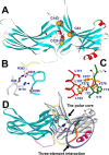
Figure 1.
Figure 1.
The three cysteines in arrestin-1 are located near the polar core. A, crystal structure of bovine arrestin-1 (PDB code 1CF1 (4)), with β strands shown in blue, α helices in red, β turns in green, and elements with no secondary structure in gray. Cys-63, Cys-128, and Cys-143 are shown as Corey-Pauling-Koltun models colored by atom (gray, carbon; blue, nitrogen; red, oxygen; yellow, sulfur). B, the polar core, consisting of Arg-175 forming a salt bridge with Asp-296, Arg-382 of the C-tail, Asp-30 of the N domain, and Asp-303, which is located on the lariat loop along with Asp-296. C, three-element interaction mediated by hydrophobic residues Val-11, Ile-12, and Phe-13 on β strand I, Leu-103, Leu-107, and Leu-111 on α helix I, and Phe-375, Val-376, and Phe-377 of the C-tail. D, the location of the polar core between the two arrestin domains and the three-element interaction in the arrestin-1 molecule is shown by circles. In B–D, the elements of the arrestin molecule are colored as follows: gray, N domain; light blue, C-domain; red, α helix I; green, β strand I.
Figure 2.
Figure 2.
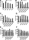
Cysteine substitution obliterates increased Rh* binding of preactivated polar core mutants. A, binding of WT bovine arrestin-1 and the indicated cysteine-less mutants. ASA, C63A, C128S, C143A; ASV, C63A, C128S, C143V; VSA, C63V, C128S, C143A; VSV, C63V, C128S, C143V. B, binding of WT arrestin-1, the preactivated R175E mutant, and the R175E mutation on the indicated cysteine-less backgrounds. C, binding of WT arrestin-1, the preactivated D296R mutant, and the D296R mutation on the indicated cysteine-less backgrounds. D, binding of WT arrestin-1, preactivated Tr arrestin-1-(1–378), and truncated mutants on the indicated cysteine-less backgrounds. E, binding of WT arrestin-1, preactivated Tr arrestin-1-(1–378), truncated arrestin-1 carrying additional K257Q and E346H mutations (QHTr), and QHTr mutants on the indicated cysteine-less backgrounds. F, the binding of WT arrestin-1, preactivated arrestin-1 with the triple substitution F375A, V376A, F377A (3A), arrestin-1–3A carrying additional K257Q and E346H mutations (QH3A), and QH3A mutants on the indicated cysteine-less backgrounds. In all panels, the columns are colored as follows: black, P-Rh*; white, Rh*. The means ± S.D. of two experiments, each performed in duplicate, are shown. The data were analyzed by one-way ANOVA (separately for P-Rh* and Rh* binding) with protein as a main factor, followed by Bonferroni-Dunn test with corrections for multiple comparisons. Statistical significance of the differences (compared with the respective base mutant with WT cysteines) is as follows: *, p < 0.05; **, p < 0.01; ***, p < 0.001.
Figure 3.
Figure 3.
Cysteine substitutions affect mouse arrestin-1 exactly like the bovine homologue. The binding of WT mouse arrestin-1 (mWT), the mASA cysteine-less mutant (C64A, C129S, C144A), as well as mutants with activating mutations (3A (L374A, V375A, P376A), Tr (truncated 1–377), D297R, and R176E) on the mWT and mASA background. The columns are colored as follows: black, P-Rh*; white, Rh*. The means ± S.D. of two experiments, each performed in duplicate, are shown. The data were analyzed (separately for P-Rh* and Rh* binding) by unpaired Student's t test, where each Cys-less mutant was compared with the corresponding mutant with WT cysteines. Statistical significance of the differences is as follows: *, p < 0.05; **, p < 0.01; ***, p < 0.001.
Figure 4.
Figure 4.
All three cysteines contribute to the reduction of Rh* binding of preactivated polar core mutants. The binding of WT bovine arrestin-1 and the indicated mutants. ASA, C63A, C128S, C143A; individual mutations are indicated. The columns are colored as in Fig. 2. The means ± S.D. of two experiments, each performed in duplicate, are shown. The data were analyzed (separately for P-Rh* and Rh* binding) by one-way ANOVA with protein as main factor, followed by Bonferroni-Dunn test with correction for multiple comparisons. The R175E and D296R mutants with cysteine substitutions were compared with the corresponding mutants with native cysteines. Statistical significance of the differences is as follows: *, p < 0.05; **, p < 0.01; ***, p < 0.001.
Figure 5.
Figure 5.
All three cysteines contribute to reduced stability of preactivated polar core mutants. A, comparison of WT arrestin-1, the Cys-less ASA mutant, and the R175E mutant. B, comparison of WT arrestin-1, the Cys-less ASA mutant, and the D296R mutant. Both panels show the binding of WT bovine arrestin-1 and the indicated mutants to P-Rh* after incubation for 15, 30, and 60 min at 39 °C; controls (0 min) were kept on ice. The means ± S.D. of two independent experiments performed in duplicate are shown. The data were analyzed by repeated measures ANOVA with protein as main factor and time as a repeated measure factor. Statistical significance of the differences in the survival time course compared with the base R175E (A) or D296R (B) mutant is indicated by $.
Figure 6.
Figure 6.
Cysteine substitutions increase the activation energy of the polar core mutant to a greater extent than the C-terminal mutant. The binding of WT bovine arrestin-1 and the indicated mutants to P-Rh* at physiological (37 °C, black columns) and very low (0 °C, white columns) temperature. The means ± S.D. of two independent experiments performed in duplicate are shown. The data for each group (WT, Tr, and R175E) and each temperature were analyzed separately by ANOVA with protein as main factor compared with the corresponding protein with WT cysteines at the same temperature. Statistical significance of the differences is as follows: *, p < 0.05; **, p < 0.01.
Figure 7.
Figure 7.
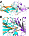
Structural basis of the function of native arrestin-1 cysteines. A, superposition of the structures of basal bovine arrestin-1 (PDB code 1CF1, molecule C (4)) and rhodopsin-bound mouse arrestin-1 (PDB code 4ZWJ (25)), with the three cysteines shown as stick models. Here and in B, the molecules are colored as follows: basal state N domain, gray; C domain, cyan; rhodopsin-bound state N domain, purple; C domain, brown. B, interdomain interface in arrestin-1 (enlargement of the center part of A). Bovine cysteine residue numbers are shown (mouse arrestin-1 numbers are n+1). Arrows indicate the shift (reflecting binding-induced rotation of the N and C domains relative to each other) that opens up the cleft between the loops in the two domains for the interaction with the intracellular part of the receptor.

References

    1. Gurevich E. V., and Gurevich V. V. (2006) Arrestins are ubiquitous regulators of cellular signaling pathways. Genome Biol. 7, 236. - PMC - PubMed
    1. Sullivan L. S., Bowne S. J., Koboldt D. C., Cadena E. L., Heckenlively J. R., Branham K. E., Birch D. G., Wheaton D. H., Jones K. D., Ruiz R. S., Pennesi M. E., Yang P., Davis-Boozer D., Northrup H., Gurevich V. V., et al. (2017) A novel dominant mutation in SAG, the Arrestin-1 gene, is a common cause of retinitis pigmentosa in Hispanic families in the southwestern United States. Invest. Ophthalmol. Vis. Sci. 58, 2774–2784 - PMC - PubMed
    1. Granzin J., Wilden U., Choe H. W., Labahn J., Krafft B., and Büldt G. (1998) X-ray crystal structure of arrestin from bovine rod outer segments. Nature 391, 918–921 - PubMed
    1. Hirsch J. A., Schubert C., Gurevich V. V., and Sigler P. B. (1999) The 2.8 A crystal structure of visual arrestin: a model for arrestin's regulation. Cell 97, 257–269 - PubMed
    1. Han M., Gurevich V. V., Vishnivetskiy S. A., Sigler P. B., and Schubert C. (2001) Crystal structure of β-arrestin at 1.9 A: possible mechanism of receptor binding and membrane translocation. Structure 9, 869–880 - PubMed

Publication types