. 2017 Jul 7;292(27):11154-11164.
doi: 10.1074/jbc.M116.766527. Epub 2017 May 23.

Structural and enzymatic insights into species-specific resistance to schistosome parasite drug therapy

Affiliations

Structural and enzymatic insights into species-specific resistance to schistosome parasite drug therapy

Alexander B Taylor et al. J Biol Chem. .

Abstract

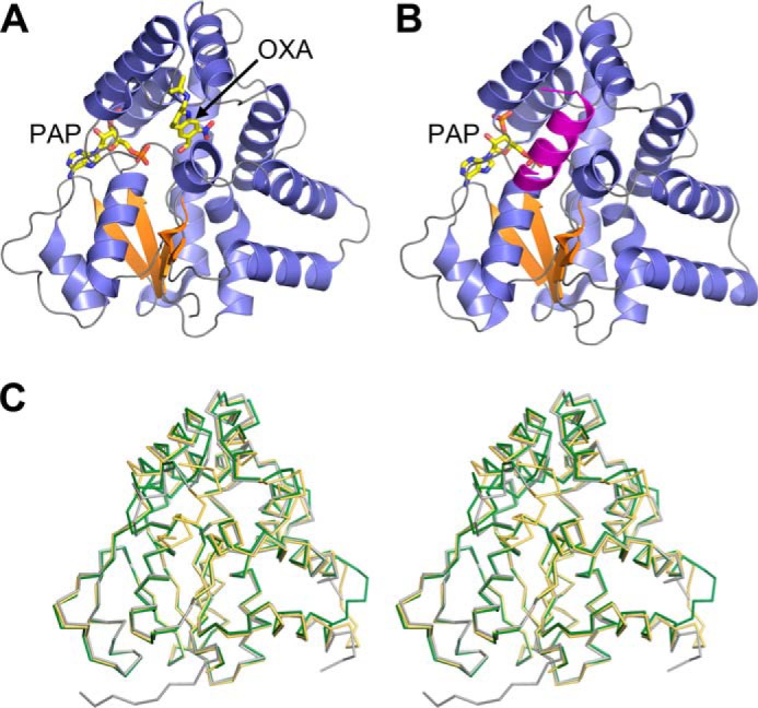
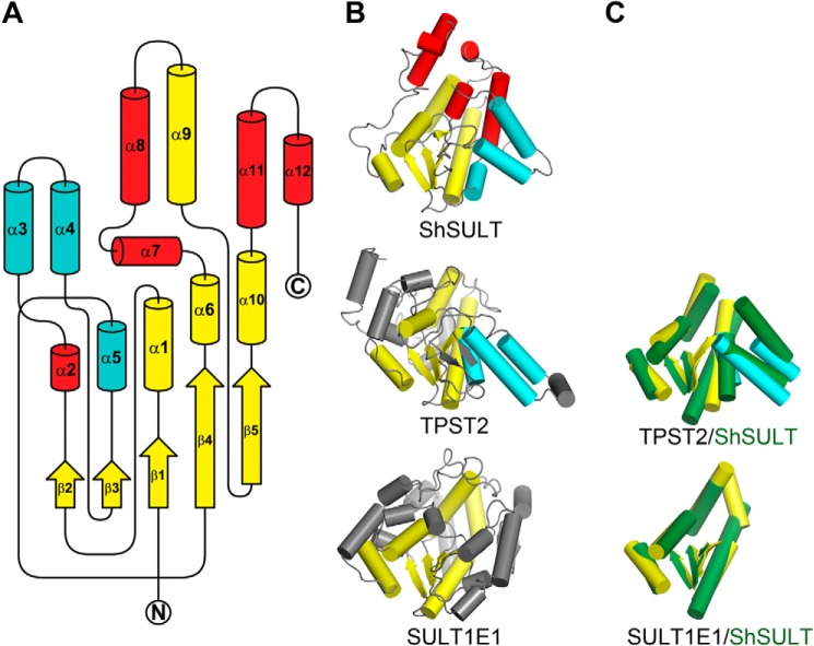
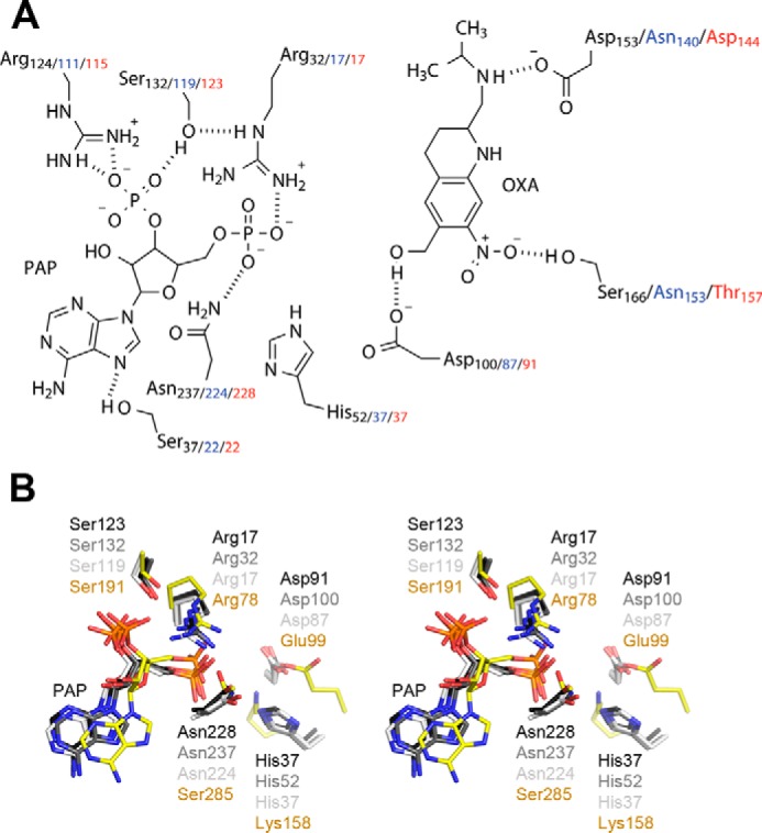
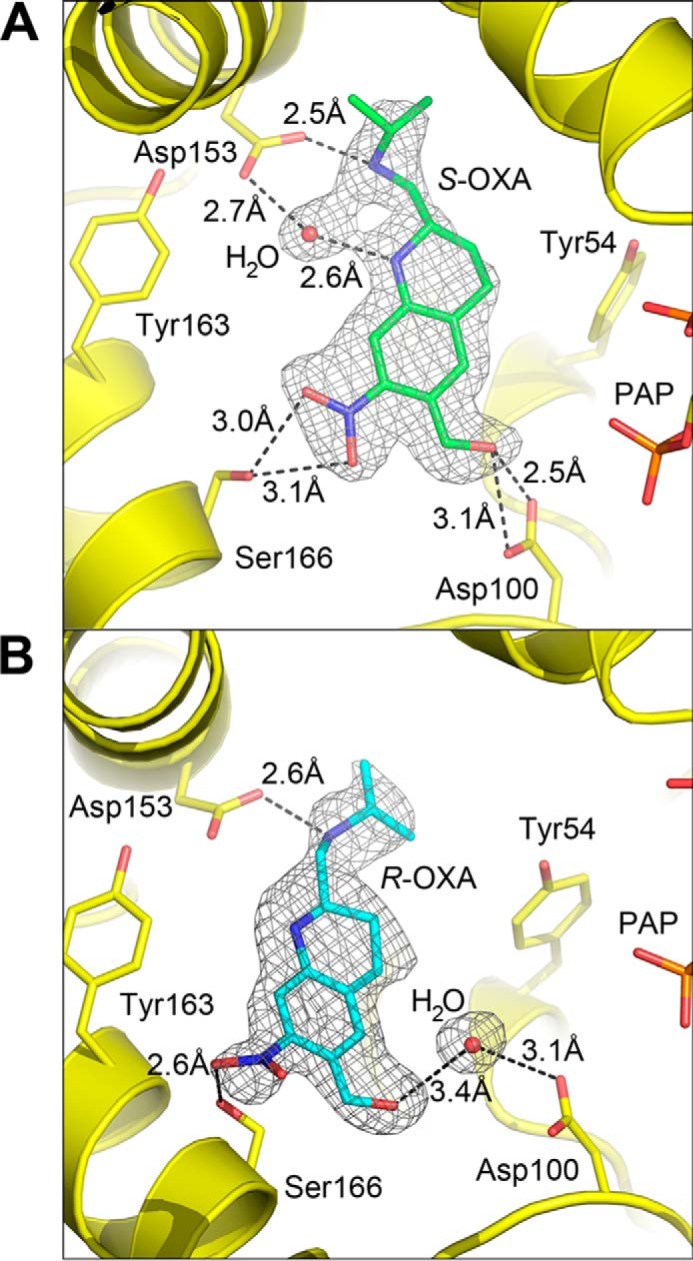
The antischistosomal prodrug oxamniquine is activated by a sulfotransferase (SULT) in the parasitic flatworm Schistosoma mansoni. Of the three main human schistosome species, only S. mansoni is sensitive to oxamniquine therapy despite the presence of SULT orthologs in Schistosoma hematobium and Schistosoma japonicum The reason for this species-specific drug action has remained a mystery for decades. Here we present the crystal structures of S. hematobium and S. japonicum SULTs, including S. hematobium SULT in complex with oxamniquine. We also examined the activity of the three enzymes in vitro; surprisingly, all three are active toward oxamniquine, yet we observed differences in catalytic efficiency that implicate kinetics as the determinant for species-specific toxicity. These results provide guidance for designing oxamniquine derivatives to treat infection caused by all species of schistosome to combat emerging resistance to current therapy.

Keywords: drug resistance; enzyme kinetics; enzyme structure; helminth; oxamniquine; parasite; schistosome; schistosomiasis; structural biology; sulfotransferase.


Conflict of interest statement

The authors declare that they have no conflicts of interest with the contents of this article

Figures

Figure 1.
Figure 1.
A, schematic representation of the ShSULT–PAP–oxamniquine (OXA) complex crystal structure. β strands are colored orange, and α helices are colored slate blue. B, schematic representation of the SjSULT–PAP complex crystal structure. Note that the predicted biological assembly is shown with a chain-swapped C-terminal α helix from the crystal symmetry-related molecule colored magenta. For clarity, the C-terminal Gly-Ser His8 affinity tag is not shown. C, walleyed stereoview of the superposition of the Cα traces of ShSULT (dark green) and SjSULT (light orange) onto SmSULT (gray).
Figure 2.
Figure 2.
A, diagram of secondary structure topology shared between the three schistosome SULTs illustrating the variation in fold as compared with mammalian SULTs. The β strands and α helices in yellow are common between the known schistosome and mammalian SULT protein structures, including SULTs 1A1, 1A3, 1B1, 1C1, 1C2, 1C3, 1D1, 1E1, 2A1, 2A3, and 4A1 and TPST2. Additionally, the α helices in cyan are common between schistosome SULT and TPST2, but α helices in red do not share a mammalian enzyme counterpart. N and C termini are labeled. B, diagram of ShSULT, TPST2 (PDB code 3AP1), and SULT1E1 (PDB code 1G3M) for comparison. The color scheme of the secondary structure elements pictured is as in A. Secondary structures of TPST2 and SULT1E1 without a schistosome counterpart are shown in gray. C, superposition of common secondary structure elements (shown in isolation) between schistosome and mammalian SULTs. Mammalian secondary structures are colored as in B, and ShSULT secondary structures are colored dark green.
Figure 3.
Figure 3.
A, schematic of the ShSULT active site (SjSULT residues in blue and SmSULT residues in red) showing oxamniquine (OXA) in position for sulfate transfer from PAPS (PAP shown). Hydrogen bonds are indicated as dotted lines. B, walleyed stereoview of the superposition of TPST2 and schistosome SULT active-site residues. SmSULT, dark gray; ShSULT, medium gray; SjSULT, light gray; TPST2, yellow.
Figure 4.
Figure 4.
A, composite omit map (2mFoDFc) calculated for the ShSULT–PAP–(S)-oxamniquine (OXA) complex contoured at 1. 0 σ. B, composite omit map (2mFoDFc) calculated for the ShSULT–PAP–(R)-oxamniquine complex contoured at 1.0 σ.
Figure 5.
Figure 5.
Crystal structure of ShSULT soaked with racemic oxamniquine. A, chain B shows a single conformation for bound (S)-oxamniquine. A composite omit map (2mFoDFc) contoured at 1.0 σ is superimposed on the atoms shown. B, oxamniquine (OXA) binds in the ShSULT active site in two positions in chain A determined by the alternate conformations of Tyr-54 and Ser-166. A composite omit map (2mFoDFc) contoured at 1.0 σ is superimposed on the atoms shown. Oxamniquine alternate confirmations and corresponding hydrogen bonding interactions are indicated in green and purple. A solvent atom is positioned between Asp-153 and oxamniquine when the alternate conformation shown in green is present (compare with A).
Figure 6.
Figure 6.
Continuous-flow mass spectra of SULT reactions after 6 s. A, mass spectrum of the reaction of 10 μm SmSULT with 20 μm oxamniquine (OXA) and 50 μm PAPS in 50 mm ethanediamine acetate, 12.5 mm ammonium acetate, 5% methanol, pH 8.0. B, mass spectrum of the reaction of 10 μm SmSULT D91A with 20 μm oxamniquine and 50 μm PAPS in 50 mm ethanediamine acetate, 12.5 mm ammonium acetate, 5% methanol, pH 8.0. C, mass spectrum of the reaction of 10 μm ShSULT with 20 μm oxamniquine and 50 μm PAPS in 50 mm ethanediamine acetate, 12.5 mm ammonium acetate, 5% methanol, pH 8.0. D, mass spectrum of the reaction of 10 μm SmSULT with 20 μm oxamniquine and 50 μm PAPS in 12.5 mm ammonium acetate, 5% methanol, pH 7.0. The spectra represent averages of 200 scans at a flow rate of 1.0 μl/min at 25 °C. Ion signals are normalized to the most intense ion. E, proposed mechanism for formation of the products of the SULT reaction.

References

    1. World Health Organization (2015) http://www.who.int/mediacentre/factsheets/fs115/en
    1. Hotez P. J., Bundy D. A. P., Beegle K., Brooker S., Drake L., de Silva N., Montresor A., Engels D., Jukes M., Chitsulo L., Chow J., Laxminarayan R., Michaud C., Bethony J., Correa-Oliveira R., et al. (2006) Helminth infections: soil-transmitted helminth infections and schistosomiasis, in Disease Control Priorities in Developing Countries (Jamison D. T., Breman J. G., Measham A. R., Alleyne G., Claeson M., Evans D. B., Jha P., Mills A., and Musgrove P., eds) pp. 467–482, 2nd Ed., The World Bank, Washington, D. C. - PubMed
    1. Centers for Disease Control and Prevention (2012) http://www.cdc.gov/parasites/schistosomiasis
    1. Greenberg R. M. (2013) New approaches for understanding mechanisms of drug resistance in schistosomes. Parasitology 140, 1534–1546 - PMC - PubMed
    1. Beck L., Favre T. C., Pieri O. S., Zani L. C., Domas G. G., and Barbosa C. S. (2001) Replacing oxamniquine by praziquantel against Schistosoma mansoni infection in a rural community from the sugar-cane zone of Northeast Brazil: an epidemiological follow-up. Mem. Inst. Oswaldo Cruz 96, (suppl.) 165–167 - PubMed

Publication types

MeSH terms

LinkOut - more resources