. 2017 May 23;7(1):2293.
doi: 10.1038/s41598-017-02570-x.

Angiotensin (1-7) ameliorates the structural and biochemical alterations of ovariectomy-induced osteoporosis in rats via activation of ACE-2/Mas receptor axis

Affiliations

Angiotensin (1-7) ameliorates the structural and biochemical alterations of ovariectomy-induced osteoporosis in rats via activation of ACE-2/Mas receptor axis

Hatem M Abuohashish et al. Sci Rep. .

Abstract

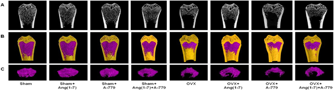
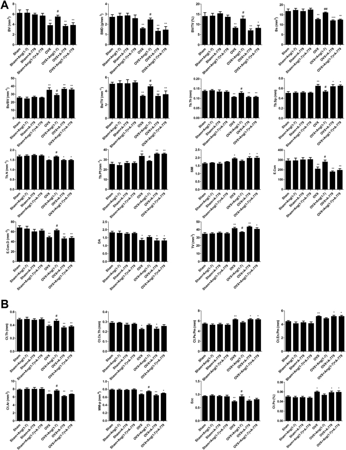
The local and systemic renin angiotensin system (RAS) influences the skeletal system micro-structure and metabolism. Studies suggested angiotensin 1-7 (Ang(1-7)) as the beneficial RAS molecule via Mas receptor activation. This study examines the function of Ang(1-7) in bone micro-architecture and metabolism in an ovariectomized (OVX) rodent model of osteoporosis. OVX rats showed structural and bone metabolic degeneration in parallel with suppressed expressions of the angiotensin converting enzyme-2 (ACE-2)/Ang(1-7)/Mas components. The infusion of Ang(1-7) markedly alleviated the altered bone metabolism and significantly enhanced both trabecular (metaphyseal) and cortical (metaphyseal-diaphyseal) morphometry. Urinary and bones minerals were also improved in OVX rats by Ang(1-7). The infusion of the heptapeptide enhanced ACE-2/Mas receptor expressions, while down-regulated AngII, ACE, and AngII type-1 receptor (AT1R) in OVX animals. Moreover, Ang(1-7) markedly improved osteoprotegerin (OPG) and lowered receptor activator NF-κB ligand (RANKL) expressions. The defensive properties of Ang(1-7) on bone metabolism, structure and minerals were considerably eradicated after blockage of Mas receptor with A-779. Ang(1-7)-induced up-regulated ACE-2/Ang(1-7)/Mas cascade and OPG expressions were abolished and the expressions of ACE/AngII/AT1R and RANKL were provoked by A-779. These findings shows for the first time the novel valuable therapeutic role of Ang(1-7) on bone health and metabolism through the ACE-2/Mas cascade.


Conflict of interest statement

The authors declare that they have no competing interests.

Figures

Figure 1
Figure 1
Effects of Ang(1-7) and/or A-779 treatments to sham and OVX animals for 6 weeks on (A) weekly body weights (BWt) increase and (B) uterus weights of Wistar albino rats. The mean weights of all OVX groups were compared to their respective sham groups in each week. The mean uterus dry weights per 100 g of final BWt of all OVX groups were compared to their respective sham groups. One-way ANOVA test followed by post hoc Student-Newman-Keuls multiple comparisons test were used for the statistical analysis. Columns and bars represent the mean ± SEM of each group (n = 8/group). Statistical significance were considered at *P < 0.05 and **P < 0.01.
Figure 2
Figure 2
Effects of Ang(1-7) and/or A-779 treatments to sham and OVX animals for 6 weeks on bone turn over biomarkers in Wistar albino rats. Levels of BALP, OC, TRACP-5b and CTX were quantified in serum, while the urinary levels of DPD was estimated using sandwich ELISA technique. One-way ANOVA test followed by post hoc Student-Newman-Keuls multiple comparisons test were used for the statistical analysis. Columns and bars represent the mean ± SEM of each group (n = 8/group). Statistical significance was considered when *P < 0.05 and **P < 0.01 as compared to Sham group and #P < 0.05 and ##P < 0.01 as compared to OVX group.
Figure 3
Figure 3
Representative (A) 2D and (B,C) 3D images of the femoral trabecular bone micro-architecture developed by micro CT. To analyze the trabecular bone micro-architecture, a volume of interest (VOI) with 1.6 mm height was selected starting 0.4 mm from the lowest end of the of the growth plate to the proximal end of the femur. (A) Sham; (B) Sham + Ang(1-7); (C) Sham + A-779; (D) Sham + Ang(1-7)+A-779; (E) OVX; (F) OVX + Ang(1-7); (G) OVX + A-779; (H) OVX + Ang(1-7)+A-779. Femur of the OVX group exhibited marked osteoporotic alterations in trabecular bone. Infusion of Ang(1-7) markedly attenuated these alterations, while A-779 abolished Ang(1-7) protective effects.
Figure 4
Figure 4
Effects of Ang(1-7) and/or A-779 treatments to sham and OVX animals for 6 weeks on distal femoral micro-architecture measured by micro-CT in Wistar albino rats. (A) Trabecular morphometric parameters in the compartment of the left distal femoral metaphyseal. (B) Cortical morphometric parameters in the metaphyseal-diaphyseal of left distal femoral bones. One-way ANOVA test followed by post hoc Student-Newman-Keuls multiple comparisons test were used for the statistical analysis. Columns and bars represent the mean ± SEM of each group (n = 8/group). Statistical significance was considered when *P < 0.05 and **P < 0.01 as compared to Sham group and #P < 0.05 and ##P < 0.01 as compared to OVX group.
Figure 5
Figure 5
Effects of Ang(1-7) and/or A-779 treatments to the sham and OVX animals for 6 weeks on the expressions of RAS different proteins and osteoclastogenesis modulating factors in the femoral bone heads. (A) Western blot analysis bands showing the expressions of AngII, Ang(1-7), AT1R, AT2R, ACE, ACE-2, MasR, RANKL and OPG. (B) Quantification of the scanning densitometry of the western blot bands (n = 8/group) expressed as arbitrary units. One-way ANOVA test followed by post hoc Student-Newman-Keuls multiple comparisons test were used for the statistical analysis. Columns and bars represent the mean ± SEM of each group. Statistical significance was considered when *P < 0.05 and **P < 0.01 as compared to Sham group and #P < 0.05 and ##P < 0.01 as compared to OVX group.

Similar articles

Cited by

References

    1. Nie, W. et al. Angiotensin-(1-7) enhances angiotensin II induced phosphorylation of ERK1/2 in mouse bone marrow-derived dendritic cells. Mol Immunol46, 355–361, doi:S0161-5890(08)00739-6 (2009). - PubMed
    1. Izu, Y. et al. Angiotensin II type 2 receptor blockade increases bone mass. J Biol Chem284, 4857–4864, doi:M807610200 (2009). - PMC - PubMed
    1. Hiruma, Y., Inoue, A., Hirose, S. & Hagiwara, H. Angiotensin II stimulates the proliferation of osteoblast-rich populations of cells from rat calvariae. Biochem Biophys Res Commun230, 176–178, doi:S0006-291X(96)95914-8 (1997). - PubMed
    1. Hatton R, Stimpel M, Chambers TJ. Angiotensin II is generated from angiotensin I by bone cells and stimulates osteoclastic bone resorption in vitro. J Endocrinol. 1997;152:5–10. doi: 10.1677/joe.0.1520005. - DOI - PubMed
    1. Schurman SJ, Bergstrom WH, Shoemaker LR, Welch TR. Angiotensin II reduces calcium uptake into bone. Pediatr Nephrol. 2004;19:33–35. doi: 10.1007/s00467-003-1361-4. - DOI - PubMed

Publication types

MeSH terms