. 2017 May 23;7(1):2295.
doi: 10.1038/s41598-017-02473-x.

TRPV4-dependent induction of a novel mammalian cold-inducible protein SRSF5 as well as CIRP and RBM3

Affiliations

TRPV4-dependent induction of a novel mammalian cold-inducible protein SRSF5 as well as CIRP and RBM3

Takanori Fujita et al. Sci Rep. .

Abstract

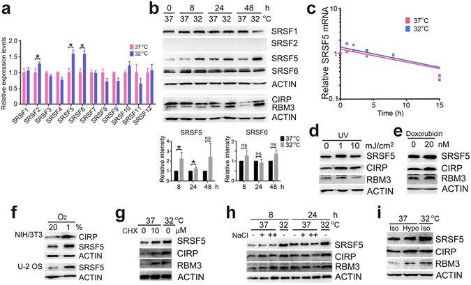
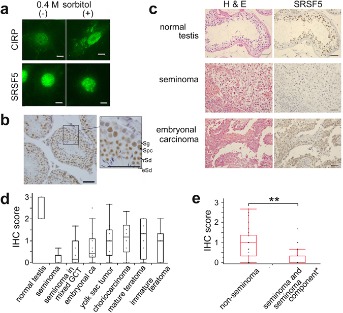
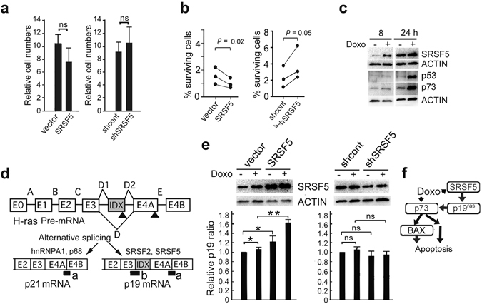
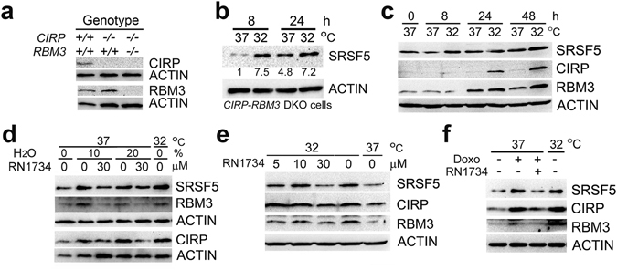
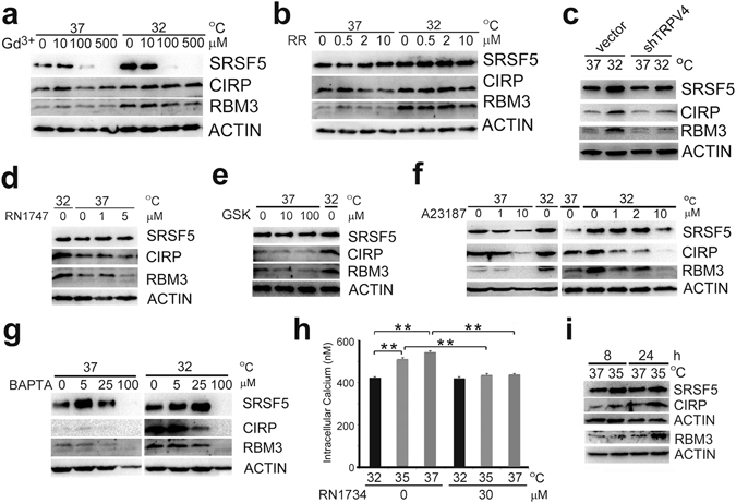
Cold-inducible RNA-binding protein (CIRP) and RNA-binding motif protein 3 (RBM3) are two evolutionarily conserved RNA-binding proteins that are structurally related to hnRNPs and upregulated in response to moderately low temperatures in mammalian cells. Although contributions of splicing efficiency, the gene promoters activated upon mild hypothermia and the transcription factor Sp1 to induction of CIRP have been reported, precise mechanisms by which hypothermia and other stresses induce the expression of mammalian cold-inducible proteins (CIPs) are poorly understood. By screening the serine/arginine-rich splicing factors (SRSFs), we report that the transcript and protein levels of SRSF5 were increased in mammalian cells cultured at 32 °C. Expression of SRSF5 as well as CIRP and RBM3 were also induced by DNA damage, hypoxia, cycloheximide and hypotonicity. Immunohistochemical studies demonstrated that SRSF5 was constitutively expressed in male germ cells and the level was decreased in human testicular germ cell tumors. SRSF5 facilitated production of p19 H-RAS, and increased sensitivity to doxorubicin in human U-2 OS cells. Induction of CIPs was dependent on transient receptor potential vanilloid 4 (TRPV4) channel protein, but seemed independent of its ion channel activity. These findings indicate a previously unappreciated role for the TRP protein in linking environmental stress to splicing.


Conflict of interest statement

The authors declare that they have no competing interests.

Figures

Figure 1
Figure 1
Induction of SRSF5 protein by various stresses. (a) Comparison of transcript levels of SRSF family members (1 to 12) in U-2 OS cells cultured at 37 °C or 32 °C for 6 h. mRNA abundance at 32 °C relative to that at 37 °C after normalization to 18S rRNA was determined for each SRSF member by quantitative RT-PCR (data indicate mean ± SEM; n = 3 per group). Statistical significance was determined by Student’s t-test. *, P < 0.05. (b) Protein levels in U-2 OS cells cultured at 37 °C or 32 °C for indicated times were analyzed by western blot (upper panels, representative results). Band intensities relative to those at 37 °C were determined after normalization to ACTIN (lower graphs, data indicate mean ± SEM; n = 3). The samples derived from the same experiment and gels/blots were processed in parallel. Statistical significance was determined by Student’s t-test. *, P < 0.05. ns, P > 0.05. (c) U-2 OS cells were cultured at 37 °C or 32 °C in the presence of 5 μg/ml actinomycin D for indicated times. SRSF5 mRNA levels relative to those at time 0 were determined by quantitative RT-PCR after normalization to 18S rRNA (data indicate mean ± SEM; n = 3). (d and e) U-2 OS cells were cultured at 37 °C for 8 h after exposure to indicated doses of UV (d) or in the presence or absence of 20 nM doxorubicin (e). Cell lysates were analyzed by western blot (representatives of 4 or 3 independent experiments). (f) NIH/3T3 and U-2 OS cells were cultured at 37 °C under normoxia (20%) or hypoxia (1% O2) for 8 h, and analyzed by western blot (representative of 4 independent experiments). (g) U-2 OS cells were cultured at 37 °C for 12 h in the presence of indicated doses of cycloheximide (CHX), and analyzed by western blot (representative of 5 independent experiments). (h) U-2 OS cells were incubated with regular media (−) or media containing additional 60 (+) or 160 (++) mM NaCl at 37 °C for 8 or 24 h, and analyzed by western blot (representative of 4 independent experiments). (i) U-2 OS cells were incubated with regular media (Iso) or hypotonic media containing 10% (volume/volume) of H2O (Hypo) at 37 °C for 8 h, and analyzed by western blot (representative of 3 independent experiments). Full-length blots are presented in Supplementary Fig. 6.
Figure 2
Figure 2
Subcellular localization and testicular expression of SRSF5 protein. (a) Localization of SRSF5 and CIRP proteins detected by immunofluorescence microscopy in NIH/3T3 cells cultured in PBS with (+) or without (−) 0.4 M sorbitol at 37 °C for 2 h. Scale bars, 50 μm. (b) Immunohistochemical (IHC) detection of SRSF5 protein in the testis of 6-wk-old C57BL/6 J mouse. Square region was enlarged in the right panel. Sg, spermatogonia; Spc, spermatocytes; rSd, round spermatids; eSd, elongated spermatids. Scale bars, 50 μm. (c) Hematoxylin and eosin (H&E) staining and IHC staining for SRSF5 of human normal testis and testicular germ cell tumors. Representative results are shown. Scale bars, 50 μm. (d) IHC scoring of SRSF5 protein levels in human normal testes and germ cell tumors (data indicate mean ± SEM. See Methods for the numbers of samples). (e) IHC comparison of SRSF5 protein levels between non-seminomas (n = 19) and seminoma components of mixed tumors and pure seminomas (n = 30) (data indicate mean ± SEM). Statistical significance was determined by Student’s t-test. **, P < 0.01.
Figure 3
Figure 3
Effects of SRSF5 on cell proliferation. (a) Stable transfectants of U-2 OS cells overexpressing SRSF5, vector alone, shRNA control and shRNA against SRSF5 (shSRSF5) were cultured at 37 °C for 4 days. Cell numbers were determined and expressed as relative to those at day 0 (mean ± SEM; n = 3). ns, P > 0.05. (b) Transfectants were cultured at 37 °C in the presence of 100 nM doxorubicin (Doxo) for 4 days. Numbers of surviving cells were determined and compared with those of untreated cells (data indicate mean ± SEM; n = 3). (c) U-2 OS cells were cultured at 37 °C in the presence (+) or absence (−) of 100 nM Doxo for 8 or 24 h, and analyzed by western blot (representative of 4 independent experiments). (d) Scheme of the Taqman assays used to determine endogenous p19 and total (p19 and p21) H-RAS mRNA expression. cDNA regions amplified by E4A-E4B and E3-IDX primer pairs are indicated by a and b, respectively. E0 to E4B, exons of human H-RAS gene. Arrow heads, stop codons. (e) Transfectants were cultured as in (c) for 10 h, and analyzed by western blot and the Taqman assays (upper panels, representative results). Relative p19 ratio, the p19/total H-RAS mRNA ratio obtained for each sample after normalization to 18S rRNA, and expressed as relative to that of control cells cultured without Doxo (mean ± SEM; n = 3). *, P < 0.05. **, P < 0.01. ns, P > 0.05. (f) A model how SRSF5 enhances apoptosis induced by Doxo. Statistical significance was determined by Student’s t-test. Full-length blots are presented in Supplementary Fig. 6.
Figure 4
Figure 4
Regulatory mechanisms of SRSF5 induction. (a and b) Fibroblasts from wild-type (+/+) or knockout (−/−) mice were analyzed for expression of CIRP and RBM3 by western blot (representative of 2 independent experiments) (a). CIRP-RBM3 double knockout (DKO) fibroblasts were cultured at 37 °C or 32 °C for 8 or 24 h, and analyzed by western blot. Relative band intensities after normalization to ACTIN expression are shown below the panel (representative of 2 independent experiments) (b). (c) B22 cells were cultured at 37 °C or 32 °C for indicated times, and analyzed by western blot (representative of 3 independent experiments). (d) U-2 OS cells were incubated with isotonic regular media (0%) or hypotonic media containing 10 or 20% (volume/volume) of H2O at 37 °C or 32 °C in the presence of 0 or 30 μM RN1734 for 8 h, and analyzed by western blot (representative of 3 independent experiments). (e) U-2 OS cells were cultured at 37 °C or 32 °C in the presence of indicated concentrations of RN1734 for 24 h, and analyzed by western blot (representative of 3 independent experiments). (f) U-2 OS cells were cultured at 37 °C or 32 °C in the absence (−) or presence (+) of 30 μM RN1734 together with 20 nM doxorubicin (Doxo) for 8 h, and analyzed by western blot (representative of 4 independent experiments). Full-length blots are presented in Supplementary Fig. 6.
Figure 5
Figure 5
TRPV4 ion channel activity and induction of CIPs. (a and b) U-2 OS cells were cultured at 37 °C or 32 °C for 24 h in the presence of indicated doses of gadolinium chloride (Gd3+) (a) or ruthenium red (RR) (b), and analyzed by western blot (representatives of 3 independent experiments each). (c) U-2 OS transfectants transiently expressing vector alone or shRNA against TRPV4 (shTRPV4) were cultured at 37 °C or 32 °C for 16 h, and analyzed by western blot (representative of 5 independent experiments). (d,e,f and g) U-2 OS cells were cultured at 37 °C or 32 °C for 24 h in the presence of indicated doses of RN1747 (d), GSK1016790A (e), A23187 (f), or BAPTA-AM (g), and analyzed by western blot (representatives of 3 independent experiments each). (h) Quantification of intracellular Ca2+ concentrations by Fura-2 in U-2 OS cells. Cells were cultured at 37 °C, 35 °C or 32 °C for 24 h in the presence or absence of 30 μM RN1734 (data indicate mean ± SEM; n = 6). Statistical significance was determined by Student’s t-test. **, P < 0.01. (i) U-2 OS cells were cultured at 37 °C or 35 °C for indicated times, and analyzed by western blot (representative of 4 independent experiments). Full-length blots are presented in Supplementary Fig. 6.

Similar articles

Cited by

References

    1. Damann N, Voets T, Nilius B. TRPs in our senses. Curr Biol. 2008;18:R880–R889. doi: 10.1016/j.cub.2008.07.063. - DOI - PubMed
    1. Holzer P, Izzo AA. The pharmacology of TRP channels. Br J Pharmacol. 2014;171:2469–2473. doi: 10.1111/bph.12723. - DOI - PMC - PubMed
    1. Clapham DE, Julius D, Montell C, Schultz G. International Union of Pharmacology. XLIX. Nomenclature and structure-function relationships of transient receptor potential channels. Pharmacol Rev. 2005;57:427–450. doi: 10.1124/pr.57.4.6. - DOI - PubMed
    1. Benemei S, Patacchini R, Trevisani M, Geppetti P. TRP channels. Curr Opin Pharmacol. 2015;22:18–23. doi: 10.1016/j.coph.2015.02.006. - DOI - PubMed
    1. Steinberg X, Lespay-Rebolledo C, Brauchi S. A structural view of ligand-dependent activation in thermoTRP channels. Front Physiol. 2014;5:171. doi: 10.3389/fphys.2014.00171. - DOI - PMC - PubMed

Publication types

MeSH terms