. 2017 May 9:8:794.
doi: 10.3389/fmicb.2017.00794. eCollection 2017.

New Functions and Subcellular Localization Patterns of c-di-GMP Components (GGDEF Domain Proteins) in B. subtilis

Affiliations

New Functions and Subcellular Localization Patterns of c-di-GMP Components (GGDEF Domain Proteins) in B. subtilis

Patricia Bedrunka et al. Front Microbiol. .

Abstract

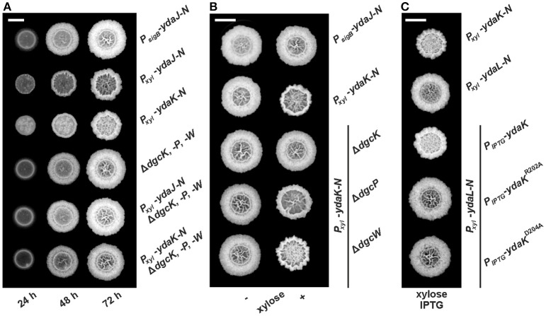
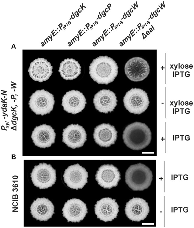
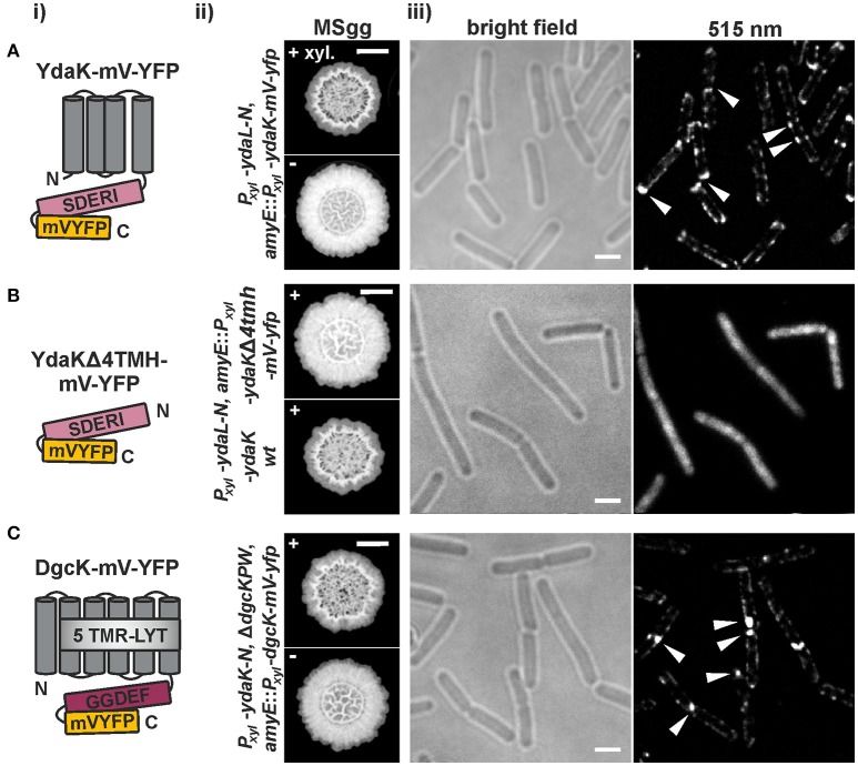
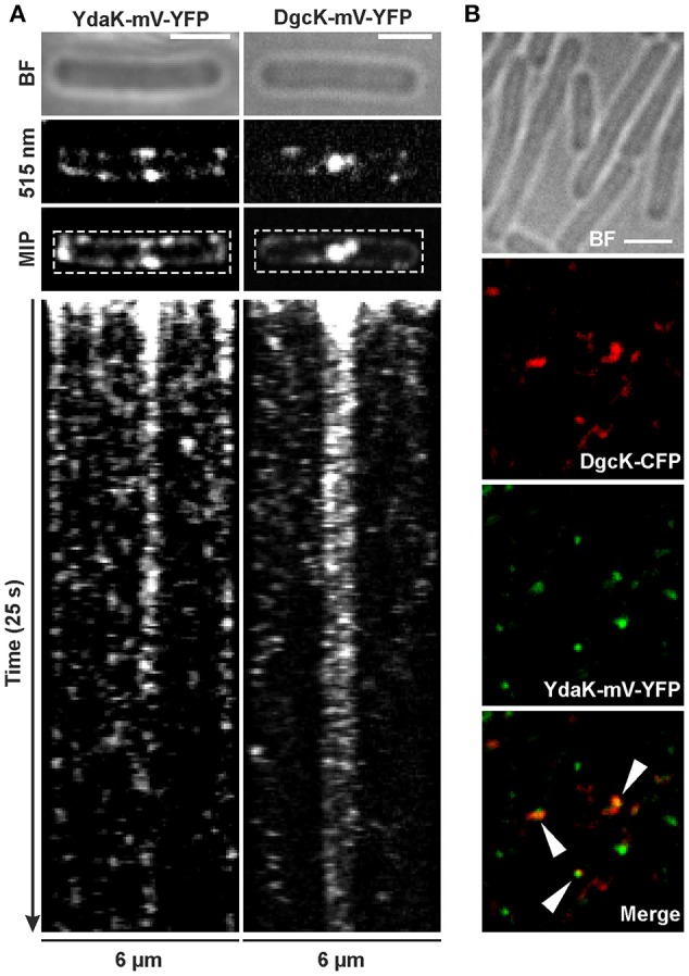
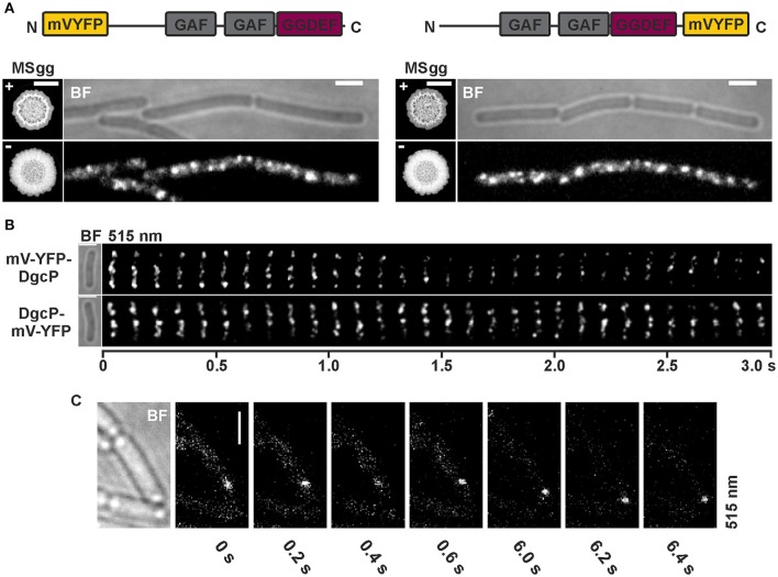
The universal and pleiotropic cyclic dinucleotide second messenger c-di-GMP is most prominently known to inversely regulate planktonic and sessile lifestyles of Gram-negative species. In the Gram-positive model organism Bacillus subtilis, intracellular c-di-GMP levels are modulated by a concise set of three diguanylate cylases (DgcK, DgcP, DgcW) and one phosphodiesterase (PdeH). Two recent studies have reported the negative influence of the c-di-GMP receptor DgrA (PilZ domain protein) on swarming motility indicating a conserved role of this second messenger across the bacterial domain. However, it has been suggested that the degenerated GGDEF protein YdaK and the inactive EAL domain protein YkuI may also function as c-di-GMP receptors regulating potentially other processes than motility. Here we describe a novel c-di-GMP dependent signaling network in B. subtilis regulating the production of an unknown exopolysaccharide (EPS) that leads to strongly altered colony morphologies upon overproduction. The network consists of the c-di-GMP receptor YdaK and the c-di-GMP synthetase DgcK. Both proteins establish a spatially close signal-effector cluster at the membrane. The cytoplasmic DgcP synthetase can complement for DgcK only upon overproduction, while the third c-di-GMP synthetase, DgcW, of B. subtilis is not part of the signaling pathway. Removal of the regulatory EAL domain from DgcW reveals a distinct function in biofilm formation. Therefore, our study is compatible with the "local pool signaling" hypothesis, but shows that in case of the yda operon, this can easily be overcome by overproduction of non-cognate DGCs, indicating that global pools can also confer signals to regulatory circuits in a Gram-positive bacterium.

Keywords: Bacillus subtilis; Biofilm formation; c-di-GMP signaling; exopolymeric substances; protein dynamics; signal transduction.


Figures

Figure 1
Figure 1
Combinatorial deletions of dgc genes and particularly inactivation of dgcK and disruption of the putative YdaK I-site motif RxxD lead to an inhibition of Yda(J)KLMN-mediated EPS production in B. subtilis. (A) Top view of B. subtilis macro colony morphology and expansion on biofilm promoting medium (MSgg, Branda et al., 2001) supplemented with 0.1% (v/v) xylose, 40 μg/ml Congo Red (CR) and 20 μg/ml Coomassie Brilliant Blue (CB) at different timepoints for WT NCIB3610 (PsigB-ydaJ-N), NCIB3610-PB53 (Pxyl-ydaJ-N), NCIB3610-PB55 (Pxyl-ydaK-N), DS1809 (ΔdgcK, -P, -W), DS1809-PB53 (ΔdgcK, -P, -W, Pxyl-ydaJ-N), and DS1809-PB55 (ΔdgcK, -P, -W, Pxyl-ydaK-N) at 25°C. Reduced colony expansion and altered wrinkle patterns (hyper-wrinkles) indicate EPS production by the products of the yda operon. (B) B. subtilis biofilm morphology on MSgg (+ CR, CB) solid medium 72 h post-inoculation in the absence and presence of 0.1% (v/v) xylose for wild type strain NCIB3610 (PsigB-ydaJ-N), the overexpression strain NCIB3610-PB55 (Pxyl-ydaK-N) and combined overexpression and deletion mutants: DS9305-PB55 (ΔdgcK; Pxyl-ydaK-N), DS9537-PB55 (ΔdgcP; Pxyl-ydaK-N), and DS9883-PB55 (ΔdgcW; Pxyl-ydaK-N). (C) Colony morphology of the overproduction mutant strains NCIB3610-PB55 (Pxyl-ydaK-N) and NCIB3610-PB56 (Pxyl-ydaL-N) and the complementation strains NCIB3610-PB56-XG003 (Pxyl-ydaL-N; amyE::PIPTG-ydaK), NCIB3610-PB56-PB80 (Pxyl-ydaL-N, amyE::PIPTG-ydaKR202A), NCIB3610-PB56-PB81 (Pxyl-ydaL-N, amyE::PIPTG-ydaKD205A) grown at 25°C on MSgg (+CR, CB) solid medium with 0.1% (v/v) xylose and 1 mM IPTG after 72 h. Bars correspond to 5 mm.
Figure 2
Figure 2
Altered biofilm formation due to ydaK-N overexpression can be restored in a dgc triple mutant by providing dgcK, dgcP, and dgcWΔeal in trans. (A) Colony development on MSgg medium supplemented with CR 40 μg/ml, CB 20 μg/ml in the presence or absence of 0.1% (v/v) xylose and/ or 1 mM IPTG, respectively, 72 h after incubation at 25°C by strains: DS1809-PB55-XG004 (ΔdgcK, -P, -W, Pxyl-ydaK-N, amyE::PIPTG-dgcK), DS1809-PB55-XG002 (ΔdgcK, -P, -W, Pxyl-ydaK-N, amyE::PIPTG-dgcP), DS1809-PB55-XG001 (ΔdgcK, -P, -W, Pxyl-ydaK-N, amyE::PIPTG-dgcW), DS1809-PB55-XG086 (ΔdgcK, -P, -W, Pxyl-ydaK-N, amyE::PIPTG- dgcWΔeal). Scale bar: 5 mm. (B) Biofilm colony morphology of B. subtilis NCIB3610 strains individually overexpressing dgcK (strain NCIB3610-XG004, amyE::PIPTG-dgcK), dgcP (NCIB3610-XG002, amyE::PIPTG-dgcP), dgcW (NCIB3610-XG001, amyE::PIPTG-dgcW), and dgcWΔeal (NCIB3610-XG086, amyE::PIPTG-dgcWΔeal) from the ectopic amylase locus. Scale bars: 5 mm.
Figure 3
Figure 3
Functional translational mV-YFP-fusions of the c-di-GMP receptor YdaK and of the synthase DgcK form subcellular assemblies at the cell poles and septa of exponentially growing B. subtilis NCIB3610 (i) Schematic representation of (A) YdaK-mV-YFP-, (B) YdaKΔ4TMH-mV-YFP- and of (C) DgcK-mV-YFP- domain organization and topology (predicted by SMART). Gray, TM helices; light gray, predicted TM-receptor domain 5TMR–LYT; purple, GGDEF domain; light purple, GGDEF domain harboring the degenerated active site motif SDERI; yellow, C-terminal mV-YFP. (ii) Verification of functionality of (A) YdaK-mV-YFP (strain: NCIB3610-PB56-PB57; Pxyl-ydaL-N, amyE::Pxyl-ydaK-mV-yfp) and of (C) DgcK-mV-YFP (strain: DS1809-PB55-PB90; ΔdgcK, -P, -W, Pxyl-ydaK-N, amyE::Pxyl-dgcK-mV-yfp). (B) Colony morphology of strain NCIB3610-PB56-PB100 (upper panel, Pxyl-ydaL-N, amyE::Pxyl-ydaKΔ4tmh-mV-yfp) and NCIB3610-PB56-PB16 (lower panel, Pxyl-ydaL-N, amyE::Pxyl-ydaK) in the presence of 0.1% (v/v) xylose. Altered colony morphology in the presence of xylose reflects EPS production by the products of the yda operon and functionality of the corresponding fusion proteins. Unaltered colony morphology of strain NCIB3610-PB56-PB100 in contrast to strain NCIB3610-PB56-PB16 (lower panel, Pxyl-ydaL-N, amyE::Pxyl-ydaK) reflects inability of YdaKΔ4TMH-mV-YFP to stimulate EPS production. Scale bars: 5 mm. (iii) Mid-exponential-phase B. subtilis NCIB3610 cells expressing (A) ydaK-mV-yfp (strain: NCIB3610-PB56-PB57; Pxyl-ydaL-N, amyE::Pxyl-ydaK-mV-yfp), (B) ydaKΔ4tmh-mV-yfp (strain NCIB3610-PB56-PB100; Pxyl-ydaL-N, amyE::Pxyl-ydaKΔ4tmh-mV-yfp) and (C) dgcK-mV-yfp (strain NCIB3610-PB90, amyE::Pxyl-dgcK-mV-yfp) from the amylase locus, 45 min after induction with 0.1% xylose. Bars: 2 μm. White triangles indicate subcellular clustering of YdaK-mV-YFP and DgcK-mV-YFP respectively.
Figure 4
Figure 4
Dynamics and simultaneous localization of YdaK and DgcK in B. subtilis NCIB3610. (A) Representative time-lapse kymographs of YdaK-mV-YFP (left panel, strain NCIB3610-PB57; amyE::Pxyl-ydaK-mV-yfp) and DgcK-mV-YFP (right panel, strain NCIB3610-PB90; amyE::Pxyl-dgcK-mV-yfp) 45 min after induction with 0.1% xylose (v/v). BF: bright field (first row); snapshots (second row) and maximum intensity projection (MIP, third row) from time-lapse microscopy; fourth row: kymographs of fluorescence intensities along the rectangular selection depicted in the third row. Images were taken every 0.1 s upon continous illumination with 515 nm. (B) Co-localization of DgcK-CFP (445 nm, false colored red) originated from the ectopic amyE locus and YdaK-mV-YFP (515 nm, false-colored green) produced from the original locus, triangles indicate co-localization events (strain NCIB3610-PB37-PB10; amyE::Pxyl-dgcK-cfp, PydaK-ydaK-mV-yfp). Scale bars: 2 μm.
Figure 5
Figure 5
Subcellular localization and dynamics of DgcP in B. subtilis NCIB3610. (A) Epifluorescence of cells overexpressing mV-yfp-dgcP (left panel, strain NCIB3610-PB85; amyE::Pxyl-mV-yfp-dgcP) and dgcP-mV-yfp (right panel, strain NCIB3610-PB86; amyE::Pxyl-dgcP-mV-yfp) 45 min after induction with 0.1% (v/v) xylose (Scale bars: 2 μm). The “MSgg panel” depicts the functionality assay for the corresponding fusion protein/strain in the presence and absence of 0.1% (v/v) xylose (left: DS1809-PB55-PB85, ΔdgcK, -P, -W, Pxyl-ydaK-N, amyE::Pxyl-mV-yfp-dgcP; right: DS1809-PB55-PB86, ΔdgcK, -P, -W, Pxyl-ydaK-N, amyE::Pxyl-dgcP-mV-yfp). Scale bar: 5 mm. Color code for schematic representation of corresponding fusion protein (domain organization predicted by SMART): gray: GAF (domain found in cGMP-specific phosphodiesterases, adenylyl and guanylyl cyclases and phytochromes which often serves as a cyclic nucleotide binding domain), purple: GGDEF domain (active site motif GGEEL), yellow: N- and C-terminal mV-YFP respectively. (B) Time-lapse fluorescence microscopy of DgcP fusions produced. Images were captured every 100 ms under continious illumination (515 nm). Bars: 2 μm. (C) Time-lapse microscopy of DgcP-mV-YFP produced from the original locus (strain NCIB3610-PB08). Images were captured at the time points (seconds) indicated next to the panels at time intervals of 200 ms. Bars: 2 μm.

References

    1. Anantharaman V., Aravind L. (2003). Application of comparative genomics in the identification and analysis of novel families of membrane-associated receptors in bacteria. BMC Genomics 4:34. 10.1186/1471-2164-4-34 - DOI - PMC - PubMed
    1. Asally M., Kittisopikul M., Rue P., Du Y., Hu Z., Cagatay T., et al. . (2012). Localized cell death focuses mechanical forces during 3D patterning in a biofilm. Proc. Natl. Acad. Sci. U.S.A. 109, 18891–18896. 10.1073/pnas.1212429109 - DOI - PMC - PubMed
    1. Bedrunka P., Graumann P. L. (2017). Subcellular clustering of a putative c-di-GMP-dependent exopolysaccharide machinery affecting macro colony architecture in Bacillus subtilis. Environ. Microbiol. Rep. [Epub ahead of print]. 10.1111/1758-2229.12496 - DOI - PubMed
    1. Belas R. (2014). Biofilms, flagella, and mechanosensing of surfaces by bacteria. Trends Microbiol. 22, 517–527. 10.1016/j.tim.2014.05.002 - DOI - PubMed
    1. Branda S. S., Gonzalez-Pastor J. E., Ben-Yehuda S., Losick R., Kolter R. (2001). Fruiting body formation by Bacillus subtilis. Proc. Natl. Acad. Sci. U.S.A. 98, 11621–11626. 10.1073/pnas.191384198 - DOI - PMC - PubMed

LinkOut - more resources