. 2017 May 9:8:657.
doi: 10.3389/fpls.2017.00657. eCollection 2017.

Sediment Resuspension and Deposition on Seagrass Leaves Impedes Internal Plant Aeration and Promotes Phytotoxic H2S Intrusion

Affiliations

Sediment Resuspension and Deposition on Seagrass Leaves Impedes Internal Plant Aeration and Promotes Phytotoxic H2S Intrusion

Kasper E Brodersen et al. Front Plant Sci. .

Abstract

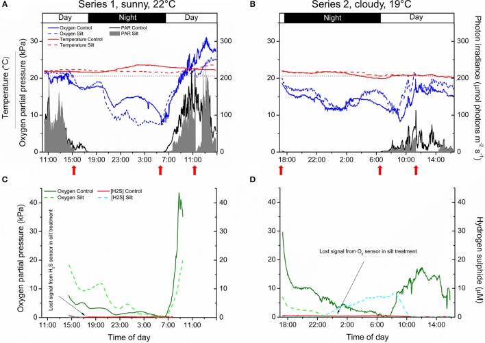
HIGHLIGHTS: Sedimentation of fine sediment particles onto seagrass leaves severely hampers the plants' performance in both light and darkness, due to inadequate internal plant aeration and intrusion of phytotoxic H2S. Anthropogenic activities leading to sediment re-suspension can have adverse effects on adjacent seagrass meadows, owing to reduced light availability and the settling of suspended particles onto seagrass leaves potentially impeding gas exchange with the surrounding water. We used microsensors to determine O2 fluxes and diffusive boundary layer (DBL) thickness on leaves of the seagrass Zostera muelleri with and without fine sediment particles, and combined these laboratory measurements with in situ microsensor measurements of tissue O2 and H2S concentrations. Net photosynthesis rates in leaves with fine sediment particles were down to ~20% of controls without particles, and the compensation photon irradiance increased from a span of 20-53 to 109-145 μmol photons m-2 s-1. An ~2.5-fold thicker DBL around leaves with fine sediment particles impeded O2 influx into the leaves during darkness. In situ leaf meristematic O2 concentrations of plants exposed to fine sediment particles were lower than in control plants and exhibited long time periods of complete meristematic anoxia during night-time. Insufficient internal aeration resulted in H2S intrusion into the leaf meristematic tissues when exposed to sediment resuspension even at relatively high night-time water-column O2 concentrations. Fine sediment particles that settle on seagrass leaves thus negatively affect internal tissue aeration and thereby the plants' resilience against H2S intrusion.

Keywords: H2S; diffusive boundary layer; dredging; in situ; microsensors; photosynthesis; seagrass; sediment.


Figures

Figure 1
Figure 1
Vertical O2 concentration profiles measured toward the leaf surface under incident photon irradiances of 0, 75, 200, and 500 μmol photons m−2 s−1. Red symbols and lines represent leaves with silt/clay-cover; black symbols and lines represent control plants, i.e., leaves without silt/clay-cover. Upper panels are measurements in water with a reduced O2 level of ~40% of air equilibrium (mimicking night-time water-column O2 conditions, approximately 8.2 kPa); Lower panels are measurements in water at 100% air equilibrium (mimicking day-time water-column O2 conditions, 20.6 kPa). Zero depth indicates the leaf surface. Symbols and error bars represent means ± SE; n = 3–4.
Figure 2
Figure 2
Vertical depth profiles of the O2 concentration measured toward the leaf surface of plants with a microbially active silt/clay-cover (red symbols and lines), with an inactivated silt/clay-cover (obtained by pre-heating the added silt/clay to 120°C in an oven for 2 h; blue symbols and lines), and without silt/clay-cover (control plants; black symbols and lines). All measurements were performed in darkness. Zero depth indicates the leaf surface. The effective DBL thickness can be estimated by extrapolating the linear O2 concentration gradient until it intersects with the constant O2 concentration in the overlying water. The distance from this point into the leaf tissue surface is a measure of the effective DBL thickness (Jørgensen and Revsbech, 1985). Symbols and error bars represent means ± SE; n = 4.
Figure 3
Figure 3
Apparent net photosynthesis rates as a function of downwelling photon irradiance (PAR, 400–700 nm) of plants with leaf silt/clay-cover (red symbols and lines) and without leaf silt/clay-cover (control plants; black symbols and lines). Rates were calculated for incident photon irradiances of 0, 75, 200, and 500 μmol photons m−2 s−1 and were fitted with an exponential function (Webb et al., 1974) with an added term to account for respiration (Spilling et al., 2010) (R40% AE, control2 = 0.93; R40% AE, silt/clay-cover2 = 0.98; R100% AE, control2 = 0.99; R100% AE, silt/clay-cover2 = 0.99). The upper panel represents measurements in water kept at 40% air equilibrium, while the lower panel represents measurements in water kept at 100% air equilibrium. Error bars are ± SE; n = 3–4.
Figure 4
Figure 4
In situ measurements of diel changes in the O2 concentration and temperature of the water-column (A,B), the light availability at leaf canopy height (A,B), and of the O2 partial pressure and H2S concentration in the meristematic tissue of Zostera muelleri plants with and without leaf silt/clay-cover, respectively (C,D) from Narrabeen Lagoon, NSW, Australia. The O2 and H2S microsensors were inserted into the shoot base close to the basal leaf meristem, which was buried ~2 cm into the sediment. The horizontal, dashed line in panels (A,B) corresponds to 100% atmospheric O2 partial pressure. Legends depict the physical/chemical water-column parameters (A,B) and the chemical species (C,D). Panels (A,C) are from the first measuring day “Series 1” (representing a sunny day), while panels (B,D) are from the second measuring day “Series 2” (representing a cloudy day). Red arrows show the timing of the fine sediment pulses in the silt/clay treatment. Measurements are recorded from the exact same plants and therefore represent changes in plant performance as a result of repeated exposure to sediment re-suspension and deposition of fine sediment particles on seagrass leaves. Note the lost signal from the inserted microsensors in the silt/clay treatment (C,D).
Figure 5
Figure 5
In situ intra-plant O2 status as a function of the O2 partial pressure in the surrounding water-column during night-time. The data were extracted from Figure 4 approximately 2 h after sunset. The gray lines represent a linear regression and are extrapolated to interception with the horizontal x-axis, to provide an estimate of the water-column O2 level where the meristematic tissue at the shoot base becomes anoxic (Rcontrol, Series 12 = 0.97; Rcontrol, Series 22 = 0.70; R2silt/clay-cover, Series 1 = 0.97; Rsilt/clay-cover, Series 22 = 0.94). Upper panels (A,B) are measurements from control plants (black symbols), while lower panels (C,D) are measurements from plants with a silt/clay-cover on the leaves (red symbols).
Figure 6
Figure 6
In situ intra-plant O2 status as a function of incident photon irradiance (PAR) during daytime. The data were extracted from Figure 4 at sunrise (Series 1). The intra-plant O2 evolution during the light-limiting phase of PAR were fitted with a linear function (Gray lines) (Rcontrol2 = 0.95, αcontrol = 0.14; Rsilt/clay-cover2 = 0.94, αsilt/clay-cover = 0.08). Black symbols show measurements from control plants, while red symbols show measurements from plants with a silt/clay-cover on the leaves.

References

    1. Barnabas A. D. (1996). Casparian band-like structures in the root hypodermis of some aquatic angiosperms. Aquat. Bot. 55, 217–225. 10.1016/S0304-3770(96)01072-8 - DOI
    1. Binzer T., Borum J., Pedersen O. (2005). Flow velocity affects internal oxygen conditions in the seagrass Cymodocea nodosa. Aquat. Bot. 83, 239–247. 10.1016/j.aquabot.2005.07.001 - DOI
    1. Borum J., Pedersen O., Greve T. M., Frankovich T. A., Zieman J. C., Fourqurean J. W., et al. (2005). The potential role of plant oxygen and sulphide dynamics in die-off events of the tropical seagrass, Thalassia testudinum. J. Ecol. 93, 148–158. 10.1111/j.1365-2745.2004.00943.x - DOI
    1. Borum J., Sand-Jensen K., Binzer T., Pedersen O., Greve T. M. (2006). Oxygen movement in seagrasses, in Seagrasses: Biology, Ecology and Conservation, eds Larkum A. W. D., Orth J. R., Duarte C. M. (Dordrecht Berlin: Springer; ), 255–270.
    1. Brodersen K. E., Koren K., Lichtenberg M., Kühl M. (2016). Nanoparticle-based measurements of pH and O2 dynamics in the rhizosphere of Zostera marina L.: effects of temperature elevation and light-dark transitions. Plant Cell Environ. 39, 1619–1630. 10.1111/pce.12740 - DOI - PubMed

LinkOut - more resources