. 2016 Aug 12:21:11.
doi: 10.1186/s11658-016-0016-y. eCollection 2016.

In vitro chondrogenesis of Wharton's jelly mesenchymal stem cells in hyaluronic acid-based hydrogels

Affiliations

In vitro chondrogenesis of Wharton's jelly mesenchymal stem cells in hyaluronic acid-based hydrogels

Ewelina Aleksander-Konert et al. Cell Mol Biol Lett. .

Abstract

Background: In this study, we evaluated the usefulness of two commercially available hyaluronic acid-based hydrogels, HyStem and HyStem-C, for the cultivation of Wharton's jelly mesenchymal stem cells (WJ-MSCs) and their differentiation towards chondrocytes.

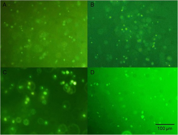
Methods: The WJ-MSCs were isolated from umbilical cord Wharton's jelly using the explant method and their immunophenotype was evaluated via flow cytometry analysis. According to the criteria established by the International Society for Cellular Therapy, they were true MSCs. We assessed the ability of the WJ-MSCs and chondrocytes to grow in three-dimensional hydrogels and their metabolic activity. Chondrogenesis of WJ-MSCs in the hydrogels was determined using alcian blue and safranin O staining and real-time PCR evaluation of gene expression in the extracellular matrixes: collagen type I, II, III and aggrecan.

Results: Chondrocytes and WJ-MSCs cultured in the HyStem and HyStem-C hydrogels adopted spherical shapes, which are characteristic for encapsulated cells. The average viability of the WJ-MSCs and chondrocytes in the HyStem hydrogels was approximately 67 % when compared with the viability in 2D culture. Alcian blue and safranin O staining revealed intensive production of proteoglycans by the cells in the HyStem hydrogels. Increased expression of collagen type II and aggrecan in the WJ-MSCs cultured in the HyStem hydrogel in the presence of chondrogenic medium showed that under these conditions, the cells have a high capacity to differentiate towards chondrocytes. The relatively high viability of WJ-MSCs and chondrocytes in both HyStem hydrogels suggests the possibility of their use for chondrogenesis.

Conlusions: The results indicate that WJ-MSCs have some degree of chondrogenic potential in HyStem and HyStem-C hydrogels, showing promise for the engineering of damaged articular cartilage.

Keywords: Chondrocytes; Chondrogenesis; HyStem; HyStem-C; Hydrogels; WJ-MSCs.


Figures

Fig. 1
Fig. 1
Cells cultured in hydrogels. a WJ-MSCs encapsulated in HyStem hydrogel. b Chondrocytes encapsulated in HyStem hydrogel. c WJ-MSCs encapsulated in HyStem-C hydrogel. d Chondrocytes encapsulated in HyStem-C hydrogel. e Attachment and spreading of WJ-MSCs on the surface of HyStem-C hydrogel. f Attachment and spreading of chondrocytes on the surface of HyStem-C hydrogel. Images taken using a Nikon Eclipse TS100 inverted microscope at a magnification of 200×; the scale bar is 100 μm
Fig. 2
Fig. 2
The viability of cells encapsulated in hydrogels. a The viability of WJ-MSCs in HyStem hydrogel after 24 h and 7 days of culture. b The viability of chondrocytes in HyStem hydrogel after 24 h and 7 days of culture. c The viability of WJ-MSCs in HyStem-C hydrogel after 24 h and 7 days of culture. d The viability of chondrocytes in HyStem-C hydrogel after 24 h and 7 days of culture. *p <0.05
Fig. 3
Fig. 3
Viability staining of cells seeded in HyStem hydrogel. a WJ-MSCs cultured in HyStem hydrogel after 7 days of culture. b WJ-MSCs cultured in HyStem hydrogel after 21 days of culture. c Chondrocytes cultured in HyStem hydrogel after 7 days of culture. d Chondrocytes cultured in HyStem hydrogel after 21 days of culture. Images taken with an Olympus BX60 at magnification 200×; the scale bar is 100 μm
Fig. 4
Fig. 4
Alcian blue staining of GAG deposition in WJ-MSC-seeded hydrogels during chondrogenesis. a WJ-MSCs cultured in HyStem hydrogel in the presence of growth medium after 21 days of culture. b WJ-MSCs cultured in HyStem hydrogel in the presence of chondrogenic medium after 21 days of culture. c WJ-MSCs cultured in HyStem-C hydrogel in the presence of growth medium after 21 days of culture. d WJ-MSCs cultured in HyStem-C hydrogel in the presence of chondrogenic medium after 21 days of culture. Images taken using a Nikon Eclipse TS100 inverted microscope at a magnification of 100×; the scale bar is 100 μm
Fig. 5
Fig. 5
Safranin O staining of GAG deposition in WJ-MSC-seeded hydrogels during chondrogenesis. a WJ-MSCs cultured in HyStem hydrogel in the presence of growth medium after 21 days of culture. b WJ-MSCs cultured in HyStem hydrogel in the presence of chondrogenic medium after 21 days of culture. c WJ-MSCs cultured in HyStem-C hydrogel in the presence of growth medium after 21 days of culture. d WJ-MSCs cultured in HyStem-C hydrogel in the presence of chondrogenic medium after 21 days of culture. Images taken using a Nikon Eclipse TS100 inverted microscope at a magnification of 200×; the scale bar is 100 μm
Fig. 6
Fig. 6
The transcriptional activity of genes (collagen type I, II and III and aggrecan) expressed as fold changes. a Expressions for WJ-MSCs cultured in HyStem hydrogel in the presence of growth medium after 21 days of culture compared to expressions for WJ-MSCs cultured in HyStem hydrogel in the presence of chondrogenic medium. b Expressions for WJ-MSCs cultured in HyStem-C hydrogel in the presence of growth medium after 21 days of culture compared to expressions for WJ-MSCs cultured in HyStem-C hydrogel in the presence of chondrogenic medium. *p <0.05

Similar articles

Cited by

References

    1. Seogjin S, Na K. Mesenchymal stem cell-based tissue engineering for chondrogenesis. J Biomed Biotechnol. 2011;2011:1–8. - PMC - PubMed
    1. Punwar S, Kham WS. Mesenchymal stem cells and articular cartilage repair: clinical studies and future direction. Open Orthop J. 2011;5:296–301. doi: 10.2174/1874325001105010296. - DOI - PMC - PubMed
    1. Sun L, Reagan MR, Kaplan DL. Role of cartilage-forming cells in regenerative medicine for cartilage repair. Orthop Res Rev. 2010;2:85–94. doi: 10.2147/ORR.S7194. - DOI - PMC - PubMed
    1. Kock L, van Donkelaar CC, Ito K. Tissue engineering of functional articular cartilage: the current status. Cell Tissue Res. 2012;347:613–627. doi: 10.1007/s00441-011-1243-1. - DOI - PMC - PubMed
    1. Troyer DL, Weiss ML. Concise review: Wharton’s jelly-derived cells are a primitive stromal cell population. Stem Cells. 2008;26:591–599. doi: 10.1634/stemcells.2007-0439. - DOI - PMC - PubMed

LinkOut - more resources