. 2016 Nov 16:21:26.
doi: 10.1186/s11658-016-0027-8. eCollection 2016.

The Characteristics Of Human Bone-Derived Cells (HBDCS) during osteogenesis in vitro

Affiliations

The Characteristics Of Human Bone-Derived Cells (HBDCS) during osteogenesis in vitro

Edyta Wrobel et al. Cell Mol Biol Lett. .

Abstract

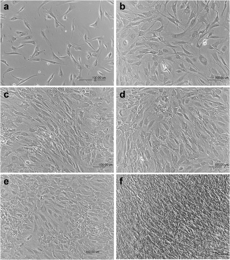
Background: The primary human bone-derived cell culture technique is used as a model to study human osteogenesis. Compared to cell line cultures, primary osteoprogenitor and osteoblast cultures provide more complex information about osteogenesis, bone remodeling and regeneration than cell line cultures.

Methods: In this study, we isolated human bone-derived cells (HBDCs) and promoted their differentiation into osteoblasts. The following parameters were evaluated: cell number and viability, total protein expression, alkaline phosphatase activity, collagenous matrix production and osteogenic genes expression, i.e., gene coding for type I collagen and alkaline phosphatase.

Results: It was proved the results show that HBDCs intensively proliferate during the first 7 days of culture followed by differentiation accompanied by an increase in alkaline phosphatase activity. Moreover, it was observed that during the differentiation of HBDCs, the expression of integrin β1 increased.

Conclusions: The process was also accompanied by changes in cell shape and rearrangement of the actin cytoskeleton and focal contacts containing FAK and the integrin β1 subunit. We suggest that the β1 integrin subunit may be a suitable new target in studies of the differentiation of primary human osteoblasts in culture.

Keywords: Human bone-derived cells; Integrins; Osteoblast differentiation; Osteogenesis in vitro; Osteogenic markers; Primary osteoblasts.


Figures

Fig. 1
Fig. 1
Morphological changes of HBDCs during their in vitro differentiation. The cells were cultured in differentiating medium. Phase contrast images were taken 24 h after cell seeding (a), and on day 7 (b), 9 (c), 14 (d), 17 (e) and 21 (f) of culture. The morphology of HBDCs changed from a fibroblastic-like to a cuboidal shape. Seven independent experiments were performed and representative images are shown. Scale bar: 100 μm
Fig. 2
Fig. 2
The cell number and viability (XTT assay) 24 h after cell seeding, and on days 7, 14 and 21 of culture. The results of the XTT assay are presented as the absorbance level. It was determined as a linear relationship between cell number and viability. Seven independent experiments were performed. The data are presented as means ± standard deviation. Cell number is in black; cell viability is in grey. Student’s t-test was used for statistical analyses. *p < 0.05 (considered significant)
Fig. 3
Fig. 3
Expression of osteoblast markers, i.e., type I collagen (COLI), alkaline phosphatase (ALP), osteocalcin (OC) determined using semi-quantitative RT-PCR. Analysis of the representative gels and the level of GAPDH, COLI, ALP and OC mRNA (the average optical density of bands; Odu) is presented in the charts (n = 3). Results are normalized to the housekeeping gene level (GAPDH). 0 – cell cultured under undifferentiating conditions in standard medium; 7, 14, 21 – cells cultured under osteogenic conditions in differentiating medium
Fig. 4
Fig. 4
The total protein content (a), collagen assay (b) and alkaline phosphatase (ALP) activity (c). The results of the collagen assay are presented as the absorbance level. The results from seven separate experiments are shown. The data are presented as means ± standard deviation. Student’s t-test was used for statistical analyses. *p < 0.05 (considered significant)
Fig. 5
Fig. 5
Localization of collagen during differentiation of HBDCs on days 1 (a), 7 (b), 14 (c) and 21 (d). Three independent experiments were performed and representative images are shown. Scale bar: 100 μm
Fig. 6
Fig. 6
Changes in the expression levels and localization of the β1 integrin subunit during differentiation of HBDCs. a Analysis of representative gels. The levels of GAPDH and β1 integrin subunit mRNA are shown in the charts. Results are normalized to the level of the housekeeping gene (GAPDH). 0 – cell cultured under undifferentiating conditions in standard medium; 7, 14, 21 – cells cultured under osteogenic conditions in differentiating medium. b, c, d Immunolocalization of the β1 integrin subunit on days 7 (b), 14 (c) and 21 (d) of HBDC differentiation. Three independent experiments were performed and representative images are shown. Scale bar: 100 μm. e Immunofluorescence staining for osteoblasts terminating in focal adhesion contacts as shown by FAK localization (on day 21 of HBDC culture). Focal adhesion plaques indicated by a white arrowhead. Three independent experiments were performed and representative images are shown. Scale bar: 100 μm. fh Localization of actin in differentiating HBDCs on days 7 (f), 14 (g) and 21 (h) of culture. Three independent experiments were performed and representative images are shown. Scale bar: 100 μm. i Cell adhesion to ECM proteins is mediated through integrin receptors, which are clustered within adhesive cellular structures known as focal adhesion or focal contact plaques. Focal adhesion plaques are supramolecular assemblies containing structural (talin, vinculin, tensin, actin filaments) and signaling components (FAK, Src, paxilin) regulating cell functions via integrins

Similar articles

Cited by

References

    1. Owen TA, Aronow M, Shalhoub V, Barone LM, Wilming L, Tassinari MS, Kennedy MB, Pockwinse S, Lian JB, Stein GS. Progressive development of the rat osteoblast phenotype in vitro: reciprocal relationships in expression of genes associated with osteoblast proliferation and differentiation during formation of the bone extracellular matrix. J Cell Physiol. 1990;143:420–30. doi: 10.1002/jcp.1041430304. - DOI - PubMed
    1. Igarashi M, Kamiya N, Hasegawa M, Kasuya T, Takahashi T, Takagi M. Inductive effects of dexamethasone on the gene expression of Cbfa1, Osterix and bone matrix proteins during differentiation of cultured primary rat osteoblasts. J Mol Histol. 2004;35:3–10. doi: 10.1023/B:HIJO.0000020883.33256.fe. - DOI - PubMed
    1. Gerstenfeld LC, Chipman SD, Kelly CM, Hodgens KJ, Lee DD, Landis WJ. Collagen expression, ultrastructural assembly, and mineralization in cultures of chicken embryo osteoblasts. J Cell Biol. 1988;106:979–89. doi: 10.1083/jcb.106.3.979. - DOI - PMC - PubMed
    1. Kartsogiannis V, Ng KW. Cell lines and primary cell cultures in the study of bone cell biology. Mol Cell Endocrinol. 2004;228:79–102. doi: 10.1016/j.mce.2003.06.002. - DOI - PubMed
    1. Czekanska EM, Stoddart MJ, Richards RG, Hayes JS. In search of an osteoblast cell model for in vitro research. Eur Cell Mater. 2012;24:1–17. doi: 10.22203/eCM.v024a01. - DOI - PubMed

LinkOut - more resources