

MITF and PU.1 inhibit adipogenesis of ovine primary preadipocytes by restraining C/EBPβ
- PMID: 28536633
- PMCID: PMC5415744
- DOI: 10.1186/s11658-016-0032-y
MITF and PU.1 inhibit adipogenesis of ovine primary preadipocytes by restraining C/EBPβ
Abstract
Background: PU box-binding protein (PU.1) is a master gene of hematopoietic lineage and an important specific transcription factor in osteoclast lineage. There is proof of its expression in adipose tissue, and it is known to significantly and negatively affect adipogenesis. However, it is unclear whether there are any other molecules involved in this process.
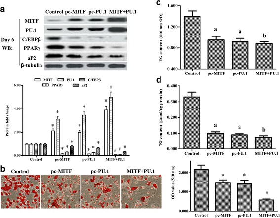
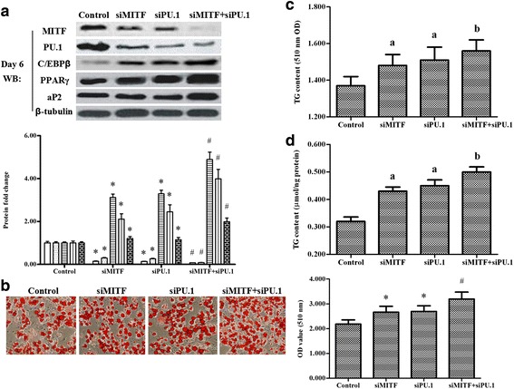
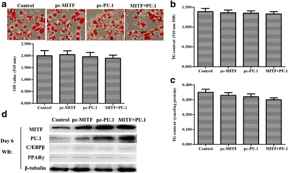
Methods: We wished to explore the effect of PU.1's co-activator microphthalmia-associated transcription factor (MITF) on the adipogenic differentiation of ovine primary preadipocytes. The expression vectors pcDNA-MITF and pcDNA-PU.1, and MITF siRNA and PU.1 siRNA were transfected or co-transfected into ovine tail primary preadipocytes. Real-time PCR and western blot analysis were applied to investigate the expression levels of PU.1 and MITF. The morphologic changes in the cells were observed under a microscope at a magnification of × 200 after staining with Oil Red O. The triglyceride (TG) content in cells was also determined after transfection.
Results: MITF and its co-activator PU.1 synergistically exhibited an opposite expression pattern to that of CCAAT-enhancer-binding protein-β (C/EBPβ) during adipogenic differentiation of ovine primary preadipocytes. Before induction of differentiation, overexpression of MITF or PU.1 inhibited the expression of C/EBPβ and adipogenesis in the cells; and knockdown of MITF or PU.1 promoted the expression of C/EBPβ and adipogenesis in the cells. The inhibitory or promotive effect was enhanced when MITF and PU.1 were co-overexpressed or co-silenced. However, when MITF and/or PU.1 were overexpressed after day 2 of differentiation, no changes in adipogenesis of the cells were observed.
Conclusions: MITF and its co-activator PU.1 inhibited adipogenesis of ovine primary preadipocytes by restraining C/EBPβ.
Keywords: Adipogenesis; CCAAT-enhancer-binding protein-β; Lineage-specific transcription factor; Microphthalmia-associated transcription factor; PU box-binding protein.
Figures

Similar articles
-
PU.1 promotes miR-191 to inhibit adipogenesis in 3T3-L1 preadipocytes.Biochem Biophys Res Commun. 2014 Aug 22;451(2):329-33. doi: 10.1016/j.bbrc.2014.07.130. Epub 2014 Aug 2. Biochem Biophys Res Commun. 2014. PMID: 25094047
-
Regulation of Wnt/β-catenin signaling by CCAAT/enhancer binding protein β during adipogenesis.Obesity (Silver Spring). 2012 Mar;20(3):482-7. doi: 10.1038/oby.2011.212. Epub 2011 Jul 14. Obesity (Silver Spring). 2012. PMID: 21760632
-
Knockdown of PU.1 AS lncRNA inhibits adipogenesis through enhancing PU.1 mRNA translation.J Cell Biochem. 2013 Nov;114(11):2500-12. doi: 10.1002/jcb.24595. J Cell Biochem. 2013. PMID: 23749759
-
Role of C/EBPβ-LAP and C/EBPβ-LIP in early adipogenic differentiation of human white adipose-derived progenitors and at later stages in immature adipocytes.Differentiation. 2013 Jan;85(1-2):20-31. doi: 10.1016/j.diff.2012.11.001. Epub 2013 Jan 11. Differentiation. 2013. PMID: 23314288
-
Knockdown of both FoxO1 and C/EBPβ promotes adipogenesis in porcine preadipocytes through feedback regulation.Cell Biol Int. 2013 Sep;37(9):905-16. doi: 10.1002/cbin.10115. Epub 2013 May 7. Cell Biol Int. 2013. PMID: 23589423
Cited by
-
PU.1 interacts with KLF7 to suppress differentiation and promote proliferation in chicken preadipocytes.Acta Biochim Biophys Sin (Shanghai). 2023 Jan 25;55(1):143-153. doi: 10.3724/abbs.2022202. Acta Biochim Biophys Sin (Shanghai). 2023. PMID: 36647727 Free PMC article.
-
Benefits of Valsartan and Amlodipine in Lipolysis through PU.1 Inhibition in Fructose-Induced Adiposity.Nutrients. 2022 Sep 12;14(18):3759. doi: 10.3390/nu14183759. Nutrients. 2022. PMID: 36145135 Free PMC article.
-
Slc25a5 regulates adipogenesis by modulating ERK signaling in OP9 cells.Cell Mol Biol Lett. 2022 Feb 2;27(1):11. doi: 10.1186/s11658-022-00314-y. Cell Mol Biol Lett. 2022. PMID: 35109789 Free PMC article.
-
miR-129 Regulates Yak Intramuscular Preadipocyte Proliferation and Differentiation through the PI3K/AKT Pathway.Int J Mol Sci. 2024 Jan 3;25(1):632. doi: 10.3390/ijms25010632. Int J Mol Sci. 2024. PMID: 38203803 Free PMC article.
-
Adipocyte PU.1 knockout promotes insulin sensitivity in HFD-fed obese mice.Sci Rep. 2019 Oct 14;9(1):14779. doi: 10.1038/s41598-019-51196-8. Sci Rep. 2019. PMID: 31611602 Free PMC article.
References
MeSH terms
Substances
LinkOut - more resources
Full Text Sources
Other Literature Sources
Molecular Biology Databases
Miscellaneous
