. 2017 Jan 25:22:3.
doi: 10.1186/s11658-017-0033-5. eCollection 2017.

MicroRNA-210 induces endothelial cell apoptosis by directly targeting PDK1 in the setting of atherosclerosis

Affiliations

MicroRNA-210 induces endothelial cell apoptosis by directly targeting PDK1 in the setting of atherosclerosis

Ying Li et al. Cell Mol Biol Lett. .

Abstract

Background: Atherosclerosis is a chronically inflammatory disease and one of the leading causes of deaths worldwide. Endothelial cell apoptosis plays a crucial role in its development. Several microRNAs (miRNAs) are reportedly involved in atherosclerotic plaque formation, including miRNA-210 (miR-210). However, the underlying mechanism of its role in endothelial cell apoptosis during atherosclerosis is still largely unknown.

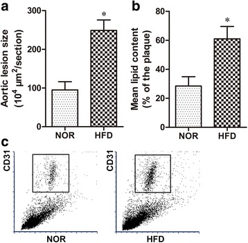
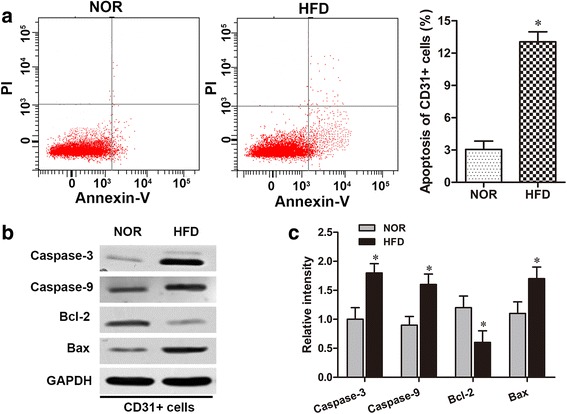
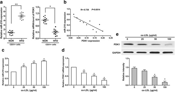
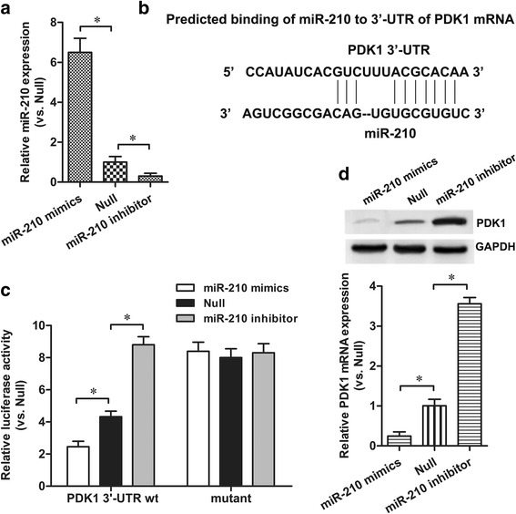
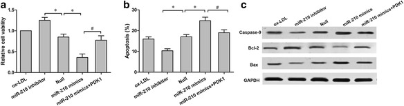
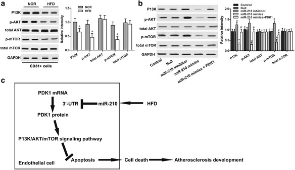
Methods: A mouse model with atherosclerosis induced by a high-fat diet (HFD) was built in ApoE (-/-) mice. The levels of endothelial cell apoptosis were determined via flow cytometry. The expressions of miR-210 and PDK1 in purified CD31+ endothelial cells from mouse aorta were measured via RT-qPCR and western blot. Binding between miR-210 and the 3'-untranslated region (UTR) of PDK1 mRNA was predicted using bioinformatics analyses and confirmed with a dual luciferase reporter assay. The effects of miR-210 were further analyzed in an in vitro model using human aortic endothelial cells (HAECs) treated with oxidized low-density lipoprotein (ox-LDL).

Results: We found that the HFD mice developed atherosclerosis in 12 weeks and had a significantly higher percentage of endothelial cell apoptosis. The upregulated level of miR-210 in the HFD mice and HAECs inversely correlated with the level of PDK1. Inhibiting miR-210 expression significantly reduced HAEC apoptosis, as evidenced by the results of the MTT and flow cytometry experiments. Further analysis identified PDK1 as the target of miR-210 and showed that PDK1 overexpression reversed the pro-apoptotic effect of miR-210 through mediation of the P13K/Akt/mTOR pathways.

Conclusion: Our study suggests a novel role for miR-210 in the progression of atherosclerosis through the regulation of endothelial apoptosis. This indicates that miR-210 might have potential in treatment of atherosclerosis.

Keywords: ApoE (-/-); Atherosclerosis; PDK1, Endothelial cell apoptosis; miR-210.


Figures

Fig. 1
Fig. 1
HFD-induced atherosclerosis in ApoE (-/-) mice after 12 weeks. a H&E staining and quantification of the plaque area of the aortic sinus. b Oil Red O staining and quantification of atherosclerotic lesions in the aortic sinus (lipid deposits stained red). c The CD31+ endothelial cells isolated from aortas using flow cytometry. n = 10 for each group. *p < 0.05
Fig. 2
Fig. 2
HFD induces endothelial cell apoptosis in ApoE (-/-) mice. a The proportions of CD31+ cell apoptosis were measured using the flow cytometry assay in the HFD and NOR groups. b The protein levels of Bax, caspase-9, caspase-3 and Bcl-2 in the CD31+ endothelial cells of the HFD and NOR groups were detected via western blot. c The protein expressions mentioned in (b) were normalized to GAPDH. *p < 0.05
Fig. 3
Fig. 3
Abnormal expressions of miR-210 and PDK1 were detected in CD31+ endothelial cells and ox-LDL-treated endothelial cells. a The expression levels of miR-210 and PDK1 in ApoE (-/-) mice were detected via RT-qPCR. b The correlation of miR-210 and PDK1 levels in ApoE (-/-) mice was measured via Peason’s coefficient correlation analysis. The effects of ox-LDL (0, 25, 50 or 100 ug/ml) on miR-210 (c) and PDK1 (d and e) expressions were detected via RT-qPCR and western blot. *p < 0.05, **p < 0.01
Fig. 4
Fig. 4
PDK1 was a target for miR-210 in HAECs. a miR-210 expression levels in HAECs with miR-210 mimics, miR-210 inhibitor or null control (Null) transfection were detected via RT-qPCR. b The binding site of miR-210 to the 3′-UTR of PDK1 was calculated through bioinformatics analysis. c Relative luciferase activities with the wild-type or mutant 3′-UTR of PDK1 were quantified. d The effect of miR-210 on PDK1 expression was examined via qRT-PCR and western blot. *p < 0.05
Fig. 5
Fig. 5
miR-210 promoted endothelial apoptosis by targeting PDK1. a HAECs cell viability was determined using the MTT assay. b Cell apoptosis of HAECs with different treatments was measured via flow cytometry. c The protein levels of Bax, caspase-9 and Bcl-2 in HAECs with different treatments were detected via western blot. *p < 0.05 vs. Null; # p < 0.05 vs. miR-210 mimics
Fig. 6
Fig. 6
MiR-210 inhibited P13K/Akt/mTOR signaling activation by targeting PDK1 in atherosclerosis. The protein levels of P13K/Akt/mTOR signaling in (a) CD31+ endothelial cells of ApoE (-/-) mice and (b) ox-LDL-treated HAECs were detected via western blot. c A schematic model of miR-210 involved in atherosclerosis-associated endothelial cell apoptosis. *p < 0.05 vs. Null; # p < 0.05 vs. miR-210 mimics

Similar articles

Cited by

References

    1. Zhang T, Tian F, Wang J, Jing J, Zhou SS, Chen YD. Endothelial cell autophagy in atherosclerosis is regulated by miR-30-mediated translational control of ATG6. Cell Physiol Biochem. 2015;37:1369–1378. doi: 10.1159/000430402. - DOI - PubMed
    1. Wang B, Zhong Y, Huang D, Li J. Macrophage autophagy regulated by miR-384-5p-mediated control of Beclin-1 plays a role in the development of atherosclerosis. Am J Transl Res. 2016;8:606–614. - PMC - PubMed
    1. Devaraj S, Jialal I. Oxidized low-density lipoprotein and atherosclerosis. Int J Clin Lab Res. 1996;26:178–184. doi: 10.1007/BF02592979. - DOI - PubMed
    1. Catapano AL, Maggi FM, Tragni E. Low density lipoprotein oxidation, antioxidants, and atherosclerosis. Curr Opin Cardiol. 2000;15:355–363. doi: 10.1097/00001573-200009000-00008. - DOI - PubMed
    1. Li K, Ching D, Luk FS, Raffai RL. Apolipoprotein E enhances microRNA-146a in monocytes and macrophages to suppress nuclear factor-kappaB-driven inflammation and atherosclerosis. Circ Res. 2015;117:e1–e11. doi: 10.1161/CIRCRESAHA.117.305844. - DOI - PMC - PubMed