. 2017 Mar 23:22:6.
doi: 10.1186/s11658-017-0037-1. eCollection 2017.

PPARγ agonist through the terminal differentiation phase is essential for adipogenic differentiation of fetal ovine preadipocytes

Affiliations

PPARγ agonist through the terminal differentiation phase is essential for adipogenic differentiation of fetal ovine preadipocytes

Yong Pu et al. Cell Mol Biol Lett. .

Abstract

Background: Although the 3T3-L1 preadipocyte cell line represents an informative model for in vitro adipogenesis research, primary cultured cells are often needed to understand particular human or animal metabolic phenotypes. As demonstrated by in vitro cultured preadipocytes from large mammalian species, primary cultured cells require specific adipogenic differentiation conditions different to that of the 3T3-L1 cell line. These conditions are also species-specific and require optimization steps. However, efficient protocols to differentiate primary preadipocytes using alternative species to rodents are scarce. Sheep represent an amenable animal model for fetal biology and developmental origins of health and disease studies. In this work, we present with the first detailed procedure to efficiently differentiate primary fetal and adult ovine preadipocytes.

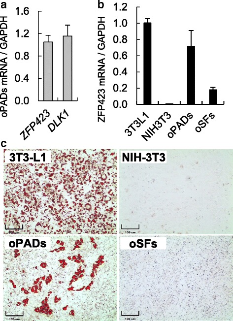
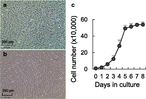
Methods: Fetal and adult ovine adipose and skin tissue harvest, preadipocyte and fibroblast isolation, proliferation, and standardization and optimization of a new adipogenic differentiation protocol. Use of commercial cell lines (3T3-L1 and NIH-3T3) for validation purposes. Oil red O stain and gene expression were used to validate adipogenic differentiation. ANOVA and Fisher's exact test were used to determine statistical significance.

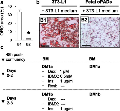
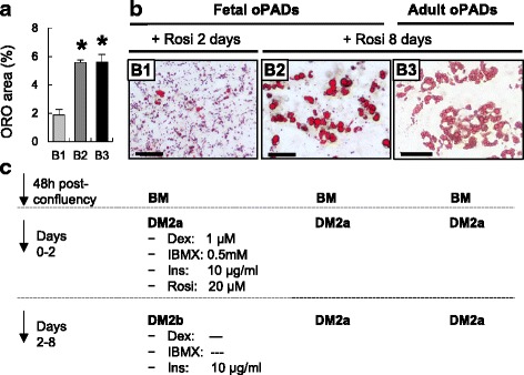
Results: Our optimized adipogenic differentiation method included a prolonged adipogenic cocktail exposure time from 2 to 8 days, higher insulin concentration, and supplementation with the peroxisome proliferator-activated receptor gamma (PPARγ) agonist, rosiglitazone. This protocol was optimized for both, fetal and adult preadipocytes.

Conclusions: Our protocol enables successful adipogenic differentiation of fetal and adult ovine preadipocytes. This work demonstrates that compared to the 3T3-L1 cell line, fetal ovine preadipocytes require a longer exposure to the differentiation cocktail, and the need for IMBX, dexamethasone, and/or the PPARγ agonist rosiglitazone through the terminal differentiation phase. They also require higher insulin concentration during differentiation to enhance lipid accumulation and similar to human primary preadipocytes, PPARγ agonist supplementation is also required for ovine adipogenic differentiation. This work highlights species-specific differences requirements for adipogenic differentiation and the need to develop standardized methods to investigate comparative adipocyte biology.

Keywords: Adipogenic differentiation; Fetal; Preadipocyte; Sheep.


Figures

Fig. 1
Fig. 1
Differentiated 3T3-L1 cells and fetal ovine preadipocytes (oPADs) with 3T3-L1 medium. Quantification of ORO positive area (mean ± SEM; a), representative ORO stain images of 3T3-L1 (B1) and fetal oPADs (B2) after 8 days of differentiation (b), and differentiation medium details (c). Scale bar: 50 μm. BM: basal medium, Dex: dexamethasone, DM: differentiation medium, Ins: insulin, oPADs: ovine preadipocytes, Rosi: rosiglitazone. Three fetal oPAD primary cells (passage 3) from three different fetuses were used. Asterisk represents significant differences (P < 0.05)
Fig. 2
Fig. 2
Effect of rosiglitazone supplementation and prolonged adipogenic cocktail exposure on ovine fetal and adult preadipocyte (oPADs) differentiation. Quantification of ORO positive area (mean ± SEM; a), representative ORO stain images of fetal (B1 (only insulin supplementation in days 3 to 8); B2 (optimized medium)) fetal and adult oPADs (B3; optimized medium) after 8 days of differentiation (b), and differentiation medium details (c). Scale bar: 50 μm. BM: basal medium, Dex: dexamethasone, DM: differentiation medium, Ins: insulin, oPADs: ovine preadipocytes, Rosi: rosiglitazone. Three fetal oPADs and three adult oPADs primary cells (passage 3) from three different fetuses and three different adult sheep, respectively, were used. Asterisks represent significant differences (P < 0.05)
Fig. 3
Fig. 3
Preadipocyte and adipogenic markers mRNA expression in murine and ovine primary cultured cells and cell lines. a) DLK1 and ZFP423 mRNA expression in ovine fetal preadipocytes (oPADs). b ZFP423 mRNA expression in undifferentiated 3T3-L1 and NIH-3T3 cell lines and fetal ovine primary cultured cells, oPADs and fetal skin fibroblasts (oSFs). c Oil red O stain of 3T3-L1 cell line (preadipocyte), NIH-3T3 cell line (fibroblast), ovine female fetal preadipocytes (oPADs), and skin fibroblasts (oSFs) after differentiation induction for 8 days. Three fetal oPADs and three fetal oSFs primary cells (passage 3) from three different fetuses were used
Fig. 4
Fig. 4
Characterization of primary fetal ovine preadipocytes (oPADs). oPADs in culture (a) and at confluency (b). c Preadipocytes growth curve (mean ± SEM) over 8 days (three fetal oPADs cell lines)
Fig. 5
Fig. 5
Dynamic gene expression in fetal ovine preadipocytes (oPADs) during differentiation. mRNA expression (mean ± SEM) through adipogenic differentiation in fetal oPADs from day 0 to 7 of differentiation when using the optimized differentiation protocol (Fig. 2b2). Different letters represent significant differences (P < 0.05) within gene and between culture days by ANOVA. Three fetal oPAD primary cells (passage 3) from three different fetuses were used. Gene expression was validated using three housekeeping genes (GAPDH, RPL27, and β-actin), but only GAPDH results are shown

Similar articles

Cited by

References

    1. Green H, Kehinde O. An established preadipose cell line and its differentiation in culture. II. Factors affecting the adipose conversion. Cell. 1975;5:19–27. doi: 10.1016/0092-8674(75)90087-2. - DOI - PubMed
    1. Ruiz-Ojeda FJ, Ruperez AI, Gomez-Llorente C, Gil A, Aguilera CM. Cell models and their application for studying adipogenic differentiation in relation to obesity: a review. Int J Mol Sci. 2016 - PMC - PubMed
    1. Huang RC, Prescott SL, Godfrey KM, Davis EA. Assessment of cardiometabolic risk in children in population studies: underpinning developmental origins of health and disease mother-offspring cohort studies. J Nutr Sci. 2015 - PMC - PubMed
    1. Benyshek DC. The “early life” origins of obesity-related health disorders: new discoveries regarding the intergenerational transmission of developmentally programmed traits in the global cardiometabolic health crisis. Am J Phys Anthropol. 2013 - PubMed
    1. Perrone S, Santacroce A, Picardi A, Buonocore G. Fetal programming and early identification of newborns at high risk of free radical-mediated diseases. World J Clin Pediatr. 2016 - PMC - PubMed