Visual rehabilitation via microperimetry in patients with geographic atrophy: a pilot study
- PMID: 28536656
- PMCID: PMC5439132
- DOI: 10.1186/s40942-017-0071-1
Visual rehabilitation via microperimetry in patients with geographic atrophy: a pilot study
Abstract
Background: Age-related macular degeneration (AMD) is the leading cause of blindness in the western world. As a consequence of AMD, patients develop structural damage that comprises the fovea and subsequently present loss of central vision, low visual acuity and unstable fixation. Contrary to what happens with anti-angiogenic treatment in neovascular AMD, there is currently no definitive treatment to reverse geographic atrophy progression. The aim of this study was to determine the effectiveness of the visual rehabilitation treatment via microperimetry in patients with geographic atrophy.
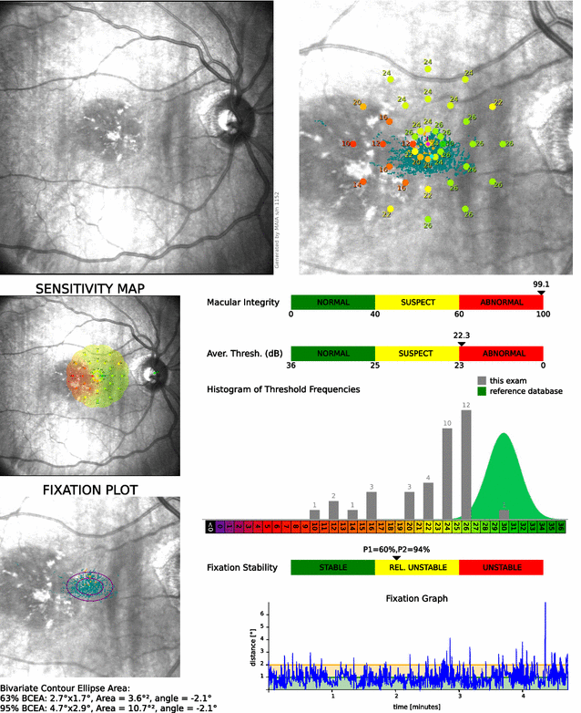
Methods: Longitudinal and prospective study, 18 patients with areas of geographic atrophy in their eye of better visual acuity were included. Macular integrity assessment (Maia) microperimeter (CentreVue, Padova, Italy) was used to diagnose retinal fixation and sensitivity in these patients. Based on these data and using the training module available in the equipment, the patients underwent visual rehabilitation sessions intended to allow the patient to establish the best possible fixation in the best area of retinal sensitivity. To determine the training effectiveness, the following variables were compared before and after: visual acuity in LogMAR scale with ETDRS charts, reading speed with Minnesota Low-Vision Reading Test (MN Read), average sensitivity threshold in microperimetry; P1 and 95% Bivariate Contour Ellipse Area (BCEA) values were used for fixation stability measurement.
Results: Mean age was 77 years old (65-92). Visual acuity of the trained eye was on average 0.7 versus 0.6 LogMAR (p = 0.006) before and one week after training. Reading speed, using both eyes, was 47 words per minute (wpm) before training and 69 wpm after training (p = 0.04). Average retinal sensitivity was 14.1 versus 14.6 db (p = 0.4). Fixation stability improved with P1 values of 45% versus 51% (p = 0.05) and 95% BCEA values of 43 versus 25 (p = 0.02) before and after training, respectively.
Conclusions: Visual training via microperimetry in patients with age-related macular degeneration is effective in improving fixation stability, reading speed, and visual acuity, measured one week after training is completed.
Keywords: Age-related macular degeneration; Fixation stability; Geographic atrophy; Microperimetry; Visual rehabilitation.
Figures


References
-
- Leibowitz HM, Krueger DE, Maunder LR, et al. The Framingham Eye Study monograph: an ophthalmological and epidemiological study of cataract, glaucoma, diabetic retinopathy, macular degeneration, and visual acuity in a general population of 2631 adults, 1973–1975. Surv Ophthalmol. 1980;24:335–610. doi: 10.1016/0039-6257(80)90015-6. - DOI - PubMed
-
- Lim JI. Age related macular degeneration. 2. New York: Informa Healthcare; 2008.
LinkOut - more resources
Full Text Sources
Other Literature Sources
Research Materials
Miscellaneous

