

Calpain-1 Expression in Triple-Negative Breast Cancer: A Potential Prognostic Factor Independent of the Proliferative/Apoptotic Index
- PMID: 28536704
- PMCID: PMC5425834
- DOI: 10.1155/2017/9290425
Calpain-1 Expression in Triple-Negative Breast Cancer: A Potential Prognostic Factor Independent of the Proliferative/Apoptotic Index
Abstract
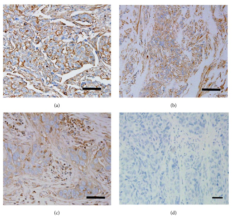
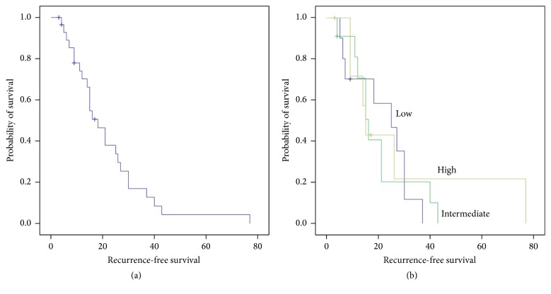
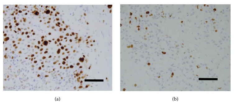
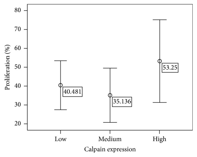
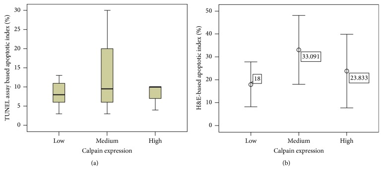
Triple-negative breast cancer (TNBC) is an aggressive type of breast cancer in which calpain system plays an important role in its cellular processes including apoptosis and proliferation. Although such roles have been assessed in tumor pathogenesis, the correlation of its expression to the proliferating/apoptotic index has not been studied yet. Immunohistochemical staining of calpain-1 was performed on paraffin-embedded tissues to correlate its expression with clinicopathological variables and outcome. The proliferation activity was determined by calculating the percentage of cells expressing the Ki-67 antigen. The apoptotic index was assessed morphologically and biochemically using Haematoxylin & Eosin method and Terminal deoxynucleotidyl transferase-mediated dUTP nick end labeling assay, respectively. Calpain-1 was significantly expressed in TNBC tissues varying from low to high with a significant correlation to lymph node status but not with the other clinicopathological variables, suggesting its role as a prognostic factor. In addition, a positive correlation was found between both apoptotic counts assays (P < 0.001, r = 0.547) as well as with proliferation (P = 0.045). Calpain-1 expression had no significant correlation with either proliferation (P = 0.29) or apoptotic indices (P = 0.071 and P = 0.100). Determining calpain-1 expression may provide relevant prognostic value for TNBC cancer patients.
Figures

Similar articles
-
Apoptosis regulator Bcl-2 is an independent prognostic marker for worse overall survival in triple-negative breast cancer patients.Int J Biol Markers. 2018 Jan;33(1):109-115. doi: 10.5301/ijbm.5000291. Int J Biol Markers. 2018. PMID: 28777433
-
P53 and Ki-67 as prognostic markers in triple-negative breast cancer patients.PLoS One. 2017 Feb 24;12(2):e0172324. doi: 10.1371/journal.pone.0172324. eCollection 2017. PLoS One. 2017. PMID: 28235003 Free PMC article.
-
Prognostic significance of proline, glutamic acid, leucine rich protein 1 (PELP1) in triple-negative breast cancer: a retrospective study on 129 cases.BMC Cancer. 2015 Oct 15;15:699. doi: 10.1186/s12885-015-1694-y. BMC Cancer. 2015. PMID: 26472563 Free PMC article.
-
Unveiling Role of MicroRNAs as Treatment Strategy and Prognostic Markers in Triple Negative Breast Cancer.Curr Pharm Biotechnol. 2020;21(15):1569-1575. doi: 10.2174/1389201021666200627201535. Curr Pharm Biotechnol. 2020. PMID: 32593278 Review.
-
Unveiling Novel Therapeutic Drug Targets and Prognostic Markers of Triple Negative Breast Cancer.Curr Cancer Drug Targets. 2021;21(11):907-918. doi: 10.2174/1568009621666210908113010. Curr Cancer Drug Targets. 2021. PMID: 34503412 Review.
Cited by
-
CAPN1 promotes malignant behavior and erlotinib resistance mediated by phosphorylation of c-Met and PIK3R2 via degrading PTPN1 in lung adenocarcinoma.Thorac Cancer. 2020 Jul;11(7):1848-1860. doi: 10.1111/1759-7714.13465. Epub 2020 May 12. Thorac Cancer. 2020. PMID: 32395869 Free PMC article.
-
Co-Expression of MHC-II and ANXA1: Mediators of PD-1/PD-L1 Therapy Resistance in Breast Cancer.Cancer Rep (Hoboken). 2025 Aug;8(8):e70291. doi: 10.1002/cnr2.70291. Cancer Rep (Hoboken). 2025. PMID: 40744683 Free PMC article.
-
Calpain-mediated Mechanoptosis in Breast Adenocarcinoma.Cancer Diagn Progn. 2023 May 3;3(3):297-301. doi: 10.21873/cdp.10215. eCollection 2023 May-Jun. Cancer Diagn Progn. 2023. PMID: 37168957 Free PMC article. Review.
-
Construction of a prognostic risk assessment model for HER2 + breast cancer based on autophagy-related genes.Breast Cancer. 2023 May;30(3):478-488. doi: 10.1007/s12282-023-01440-x. Epub 2023 Mar 1. Breast Cancer. 2023. PMID: 36856932
-
High calpain-1 expression predicts a poor clinical outcome and contributes to tumor progression in pancreatic cancer patients.Clin Transl Oncol. 2019 Jul;21(7):924-932. doi: 10.1007/s12094-018-02006-6. Epub 2018 Dec 18. Clin Transl Oncol. 2019. PMID: 30565085
References
-
- National Breast Cancer Foundation. Triple negative breast cancer. 2015, http://www.nationalbreastcancer.org/triple-negative-breast-cancer.
-
- National Breast Cancer Foundation. Metastatic breast cancer. 2015, http://www.nationalbreastcancer.org/
MeSH terms
Substances
LinkOut - more resources
Full Text Sources
Other Literature Sources
Research Materials
Miscellaneous
