. 2017 May 24:8:15221.
doi: 10.1038/ncomms15221.

Induction of resident memory T cells enhances the efficacy of cancer vaccine

Affiliations

Induction of resident memory T cells enhances the efficacy of cancer vaccine

Mevyn Nizard et al. Nat Commun. .

Abstract

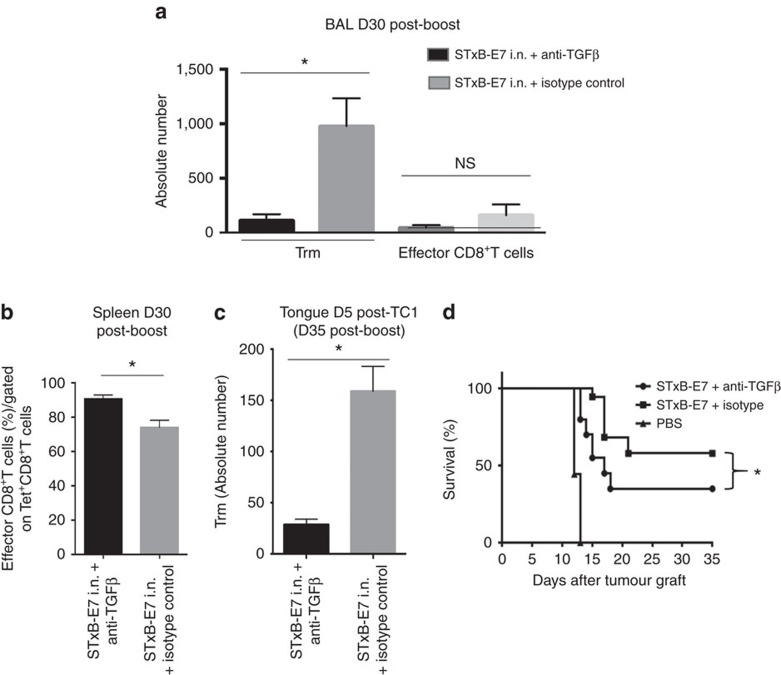
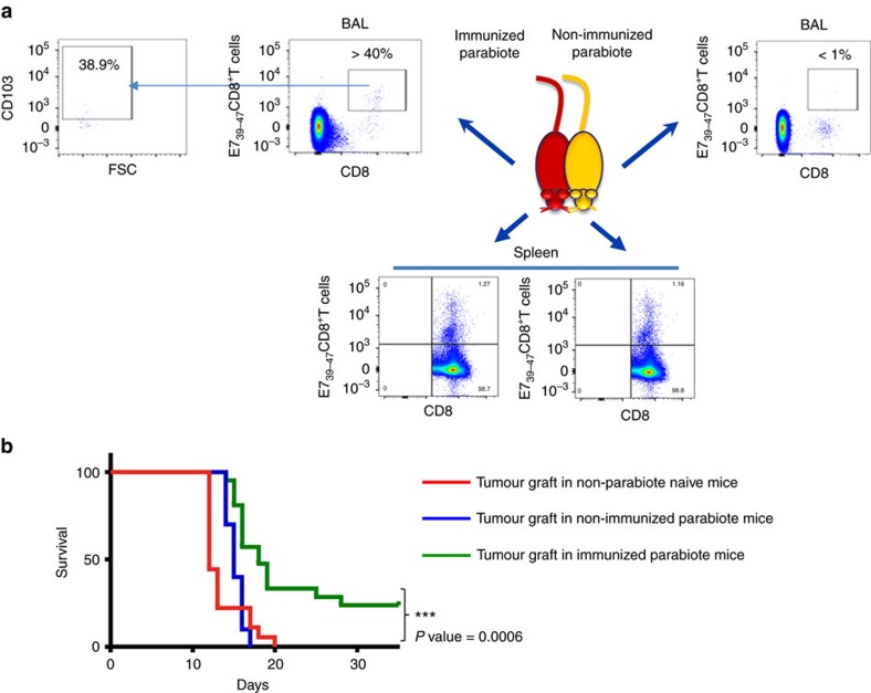
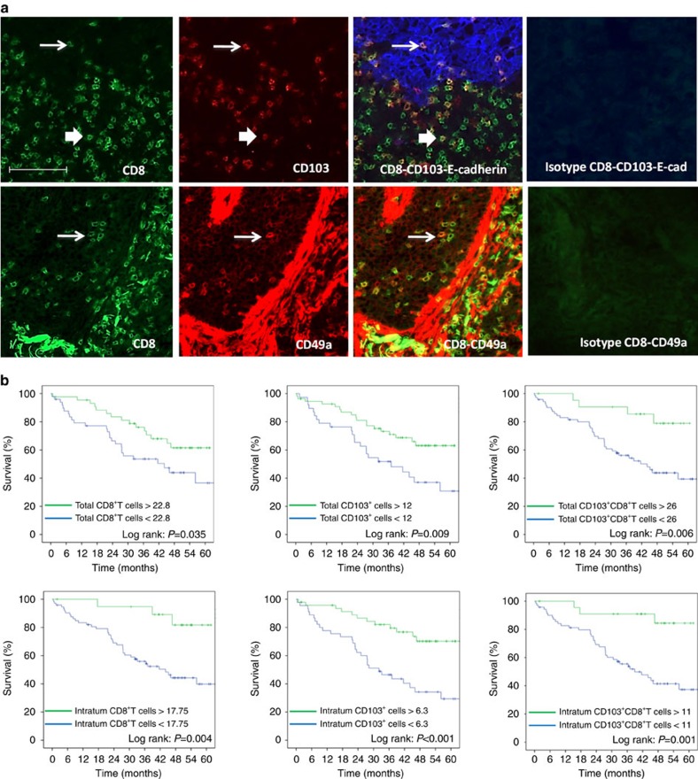
Tissue-resident memory T cells (Trm) represent a new subset of long-lived memory T cells that remain in tissue and do not recirculate. Although they are considered as early immune effectors in infectious diseases, their role in cancer immunosurveillance remains unknown. In a preclinical model of head and neck cancer, we show that intranasal vaccination with a mucosal vector, the B subunit of Shiga toxin, induces local Trm and inhibits tumour growth. As Trm do not recirculate, we demonstrate their crucial role in the efficacy of cancer vaccine with parabiosis experiments. Blockade of TFGβ decreases the induction of Trm after mucosal vaccine immunization, resulting in the lower efficacy of cancer vaccine. In order to extrapolate this role of Trm in humans, we show that the number of Trm correlates with a better overall survival in lung cancer in multivariate analysis. The induction of Trm may represent a new surrogate biomarker for the efficacy of cancer vaccine. This study also argues for the development of vaccine strategies designed to elicit them.


Conflict of interest statement

The authors declare no competing financial interests.

Figures

Figure 1
Figure 1. Heat map analysis of the gene expression pattern from antigen-specific CD8+T cells after i.n. or i.m. immunizations.
Mice were immunized via i.n. or i.m. routes (n=4 per group) with STxB-E7+α-GalCer (prime D0) and boosted at D14 with STxB-E7. At day 21, the BALs from the i.n. immunized mice and the spleens from i.m. immunized mice were collected. CD44hiTet+CD8+T cells were sorted and the RNA was extracted. Whole gene expression microarray analysis was performed according to the procedure described in the methods section. BALs CD8+T cells showed a typical Trm gene expression profile, which was not observed in splenic CD8+T cells (red square=overexpressed/blue square=underexpressed). Markers in red mean overexpressed in the CD8+T cells from the BAL, while markers in blue mean underexpressed in the CD8+T cells from the BAL. These extractions were reproduced at least three times.
Figure 2
Figure 2. Kinetics of Trm and effector CD8+T cells after i.n. immunization.
(a) Mice were i.n. immunized (prime (D0)–boost (D14)) with STxB-E7 and BALs and spleens were collected at day 21. Cells were stained and gated on living double-positive E739–47 tetramer and CD8+T cells to assess the expression of CD103 and CD49a. CD49a and CD103 double-negative cells were further stained with anti-CD62L mAb. Isotype controls were included in each experiment. (b) Absolute number of Trm cells (CD8+Tet+CD49a+CD103, CD8+Tet+CD49aCD103+, CD8+Tet+CD49a+CD103+) and effector CD8+T cells (CD8+Tet+CD49aCD103-) in BALs measured at D0, D7, D30 and D90 after a vaccine boost. (c,d) Mice were i.n. immunized or not with STxB-E7 and 7 days after the boost grafted with the TC1 tumour cells. Three and six days after the graft, the percentage (c) and the absolute number (d) of Trm and effector CD8+T cells were measured in BALs. Non-immunized mice were also included as controls. Experiments were reproduced at least three times.
Figure 3
Figure 3. Comparative functional analysis of effector CD8+T cells and Trm.
Effector CD8+T cells from BAL and spleen and Trm from BAL were sorted 7 days after the boost i.n. immunization with STxB-E7. Cells were pulsed or not with a set of concentrations of the E739–47 peptide and 24 h later IFNγ Elispot was performed. Experiments were reproduced two times and statistical analysis was performed with a two-way analysis of variance (ANOVA). Error bars indicates s.e.m. NS, not significant.
Figure 4
Figure 4. Role of Trm in the control of orthotopic tumours.
(a) Mice were i.n. immunized or not with STxB-E7 and αGalCer at D0 followed by a boost at D14 with STxB-E7. TC1 cells were grafted at D21. One day before the tumour challenge, the mice were started to be treated or not daily with FTY720 and monitored for survival. (b) Mice were i.n. immunized or not with STxB-E7 and αGalCer at D0 followed by a boost at D14 with STxB-E7. One month later, mice were grafted with TC1 cells in the tongue. Survival was monitored and represented by a Kaplan–Meier curve. Experiments were repeated at least three times. ****P<0,0001. (c,d) Mice were immunized as in a. Trm (c) and effector CD8+T cells (d) were then measured in the whole tongue 30 days after the boost without TC1 challenge (D44 after priming) for Trm or 30 days after a TC1 submucosae graft at D21 (day 51 after priming) or 30 days after the third TC1 challenge at day 111 (D141 after priming) for both Trm and effector CD8+T cells. One of two representative experiments is shown. (e) Mice were not immunized and grafted with TC1 cells at day 21 or day 51 or day 111 (red filled square). Another (green filled triangle) group of mice were (black filled nabla) immunized with STxB-E7 at D0 and D14 and grafted with TC1 at D21, D51 and D111. FTY720 was injected i.p. from (blue filled circle) D110 (one day before the third tumour challenge) until the end of the experiment. Survival was monitored. Experiments were reproduced three times. Statistical analysis for (a,b,e) used Log-rank test and for (c,d) non-parametric Mann–Whitney test. *****P<0.00001, ****P<0.0001, **P<0.01. Error bars indicates s.e.m. NS, not significant.
Figure 5
Figure 5. Modulation of Trm by TGFβ neutralization decreases cancer vaccine efficacy.
Mice were i.n. immunized (prime-boost) by STxB-E7 and treated in parallel by an anti-TGFβ or its isotype control. Treatment was stopped 1 week after the boost. Trm and effector CD8+T cells were measured 30 days after the boost in the BAL (a) and effector CD8+T cells were counted in the spleen 30 days after the boost (b). Mice were grafted with TC1 cells in the tongue 30 days after the boost, and 5 days later the number of Trm was measured in the tongue submucosae by flow cytometry to enumerate the absolute number of Trm in the presence or absence of anti-TGFβ (c). (d) Survival analysis of mice immunized by STxB-E7 combined or not with the anti-TGFβ treatment and grafted 30 days after the boost with TC1 cells. Experiments were performed with at least six mice per immunization group and repeated three times. Statistical analysis for (ac) used non-parametric Mann–Whitney test and for (d) a Log-rank test. *P<0.05. Error bars indicates s.e.m. NS, not significant.
Figure 6
Figure 6. Trm controls orthotopic tumour growth as demonstrated in parabiosis experiments.
Mice (n=5) were i.n. immunized with STxB-E7 and 7 days after the boost submitted to parabiosis surgical procedure. (a) Dot plot of T cells stained with anti-CD8 and Db-E739–47 tetramer in BALs or spleens from the immunized (left side panels) or non-immunized (right side panels) parabiont mouse 21 days after the surgery. Double-positive cells were gated for CD103 analysis from the BALs of the immunized mice (left). (b) Seven days after the parabiosis surgery, the immunized and non-immunized parabionts or naive mice (n=5 per group) were grafted in the tongue with TC1 cells and their survival was monitored. These experiments were independently reproduced three times with similar results. Statistical analysis used a Log-rank test. ***P<0.001.
Figure 7
Figure 7. Expression and prognostic value of CD103+CD8+T cells and CD49a+CD8+T in lung carcinoma patients.
(a) Top: Frozen tissue sections derived from lung carcinoma patients were stained by immunofluorescence with antibodies directed against human CD8 (green), CD103 (red) and E-cadherin (blue). The E-cadherin staining identifies the carcinomatous nests. The colocalization of CD8, CD103 and E-cadherin markers can be detected by merging the mono-staining picture. The thick arrow indicates extra-tumoural CD103+CD8+T cells; the thin arrow indicates the intratumoural CD103+CD8+T cells. Staining with isotypes controls was included for each experiment. (a) Bottom: Frozen tissue sections derived from lung carcinoma patients were stained by immunofluorescence with antibodies directed against human CD8 (green), CD49a (red). The colocalization of CD8 and CD49a markers can be detected by merging the mono-staining picture. The arrows indicate the CD49a+CD8+T cells. Staining with isotypes controls was included for each experiment (original magnification × 200). (b) Kaplan–Meier analysis of overall survival of patients with lung cancer (n=96). Tumour samples were stained for CD8 and CD103 and E-cadherin to delineate intratumoural or total infiltration (stromal and intratumoural) of cells. Patients were then divided on the basis of CD8+, CD103+ or CD8+CD103+T cell infiltration with optimized cut-off. Statistical analysis used a Log-rank test.

References

    1. Wakim L. M., Waithman J., van Rooijen N., Heath W. R. & Carbone F. R. Dendritic cell-induced memory T cell activation in nonlymphoid tissues. Science 319, 198–202 (2008). - PubMed
    1. Gebhardt T. et al. Memory T cells in nonlymphoid tissue that provide enhanced local immunity during infection with herpes simplex virus. Nat. Immunol. 10, 524–530 (2009). - PubMed
    1. Masopust D. et al. Dynamic T cell migration program provides resident memory within intestinal epithelium. J. Exp. Med. 207, 553–564 (2010). - PMC - PubMed
    1. Mackay L. K. et al. The developmental pathway for CD103(+)CD8+ tissue-resident memory T cells of skin. Nat. Immunol. 14, 1294–1301 (2013). - PubMed
    1. Beura L. K. & Masopust D. SnapShot: resident memory T cells. Cell 157, 1488–1488 e1 (2014). - PubMed

Publication types

MeSH terms