. 2017 Jul 11;8(28):45687-45697.
doi: 10.18632/oncotarget.17669.

NRAS mutations in cutaneous T cell lymphoma (CTCL) sensitize tumors towards treatment with the multikinase inhibitor Sorafenib

Affiliations

NRAS mutations in cutaneous T cell lymphoma (CTCL) sensitize tumors towards treatment with the multikinase inhibitor Sorafenib

Michael K Kießling et al. Oncotarget. .

Abstract

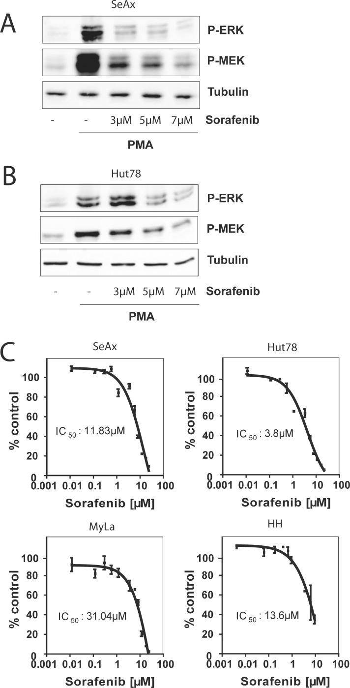
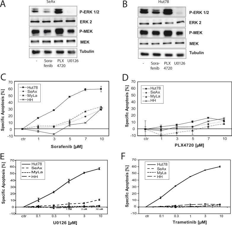
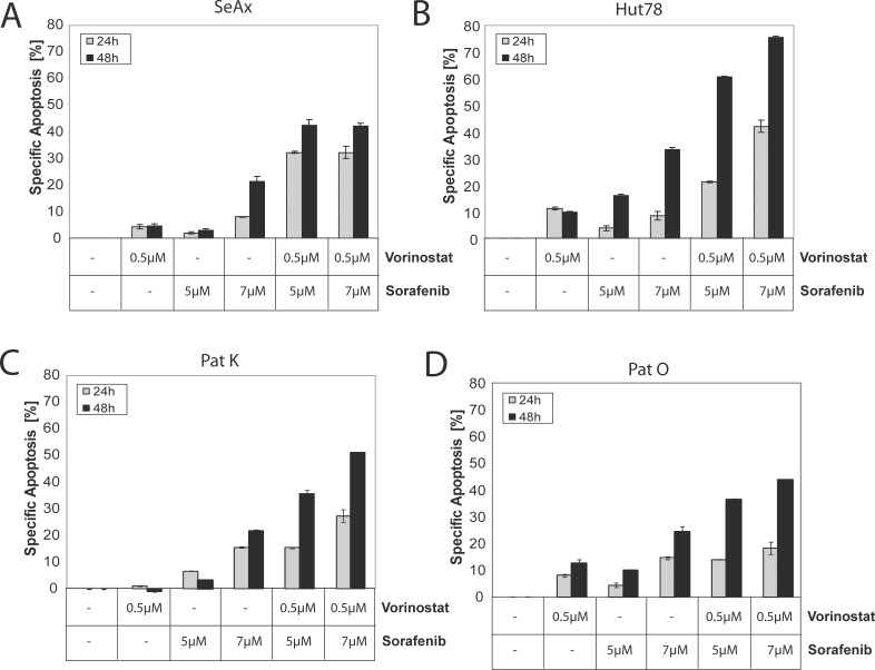
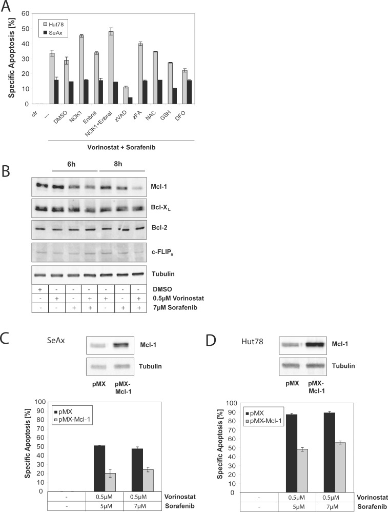
Therapy of cutaneous T cell lymphoma (CTCL) is complicated by a distinct resistance of the malignant T cells towards apoptosis that can be caused by NRAS mutations in late-stage patients. These mutations correlate with decreased overall survival, but sensitize the respective CTCL cells towards MEK-inhibition-induced apoptosis which represents a promising novel therapeutic target in CTCL. Here, we show that the multi-kinase inhibitor Sorafenib induces apoptosis in NRAS-mutated CTCL cells. CTCL cell lines and to a minor extent primary T cells from Sézary patients without NRAS mutations are also affected by Sorafenib-induced apoptosis suggesting a sensitizing role of NRAS mutations for Sorafenib-induced apoptosis. When combining Sorafenib with the established CTCL medication Vorinostat we detected an increase in cell death sensitivity in CTCL cells. The combination treatment acted synergistically in apoptosis induction in both non-mutant and mutant CTCL cells. Mechanistically, this synergistic apoptosis induction by Sorafenib and Vorinostat is based on the downregulation of the anti-apoptotic protein Mcl-1, but not of other Bcl-2 family members. Taken together, these findings suggest that Sorafenib in combination with Vorinostat represents a novel therapeutic approach for the treatment of CTCL patients.

Keywords: RAS mutation; T cell lymphoma; kinase; small molecule inhibitor; targeted therapy.


Conflict of interest statement

CONFLICTS OF INTEREST

J.P.N. received travel and congress participation funding by TEVA as well as consulting fees by TEVA and Biogen. C.D.K. received travel support for scientific conferences and lecture fees for scientific presentations from TEVA/Cephalon Pharma GmbH and Therakos, Johnson & Johnson Medical GmbH. He was a member of the TEVA Cutaneous Lymphoma Advisory Board. P.H.K. and K.G. received consulting fees by the Biogen. The project was supported by the DKFZ / Bayer HealthCare Alliance.

Figures

Figure 1
Figure 1. Sorafenib blocks RAS signaling and inhibits cell growth
Cells were left untreated, stimulated with PMA, or pre-treated with 3 μM, 5 μM, and 7μ M of Sorafenib for 30 min and then stimulated with PMA. Then, cells were lysed and the phosphorylation level of ERK and MEK was assessed by Western blot with specific anti-phospho-ERK and with specific anti-phospho-MEK antibodies. Equal loading was verified by α-tubulin. (A) Representative Western blot of SeAx cells. (B) Representative Western blot of Hut78 cells. (C) CTCL cell lines were incubated with indicated concentrations of the pan-RAF inhibitor Sorafenib for 72 hours. Cell growth was measured by Cell Titer Glo according to manufactor´s instructions. The IC50 value represents the Sorafenib concentration that inhibits 50% cell growth compared to DMSO treated control cells. The IC50 was calculated by GraphPad Prism software (San Diego, CA).
Figure 2
Figure 2. NRASQ61K sensitizes towards treatment with Sorafenib
Cells were left untreated or treated with 10 μM of Sorafenib, 3 μM of PLX4720, or 1μM of U0126 for 4 hours. Then, cells were lysed and the basal phosphorylation level of ERK and MEK was assessed by Western blot with specific anti-phospho-ERK and with specific anti-phospho-MEK antibodies. Equal loading was verified by α-tubulin, total ERK and total Mek. (A) Representative Western blot of SeAx cells. (B) Representative Western blot of Hut78 cells. (CF) All four CTCL cell lines were incubated with indicated concentrations of the used inhibitors for 48 hours. Then, apoptosis was determined and specific apoptosis was calculated according to the description in materials and methods. Data shown is representative for at least three independent experiments. (C) Cells were treated with the pan-RAF inhibitor Sorafenib. (D) Cells were incubated with indicated concentrations of the BRAF specific inhibitor PLX4720. (E, F) Similar to (C), however, all four CTCL cell lines were incubated with indicated concentrations of the MEK inhibitor U0126 (E) or Trametinib (F) for 48 h hours. Data shown is representative for two independent experiments.
Figure 3
Figure 3. Concurrent treatment with Sorafenib and Vorinostat synergistically induce apoptosis in non-mutant and in mutant cells
Cells were left untreated, treated with either Vorinostat or Sorafenib alone, or treated with different combinations of Vorinostat and Sorafenib for 24 and 48 hours. Then, apoptosis was determined and specific apoptosis was calculated according to the description in materials and methods. (A) SeAx were used. (B) Hut78 cells were used. (C, D) Same as (A, B), but CD4+ T cells isolated from two different patients were used.
Figure 4
Figure 4. Sorafenib and Vorinostat induce apoptosis synergistically via down-regulation of Mcl-1
(A) Hut78 or SeAx cells treated with either 0.5 μM Vorinostat and 5 μM Sorafenib alone, or co-treated with 1% DMSO, 1% NOK 1, 25 μM Enbrel®, 20 μM zVAD, 50 μM zFA, 20 mM N-acetyl-cysteine (NAC), 0.5 mM glutathione monoethyl ester (GSH), or 20 μM desferrioxamine (DFO) for 24 and 48 hours. Then, apoptosis was determined by flow cytometry and specific apoptosis was calculated according to the description in materials and methods. (B) SeAx cells were left untreated, treated with either Vorinostat or Sorafenib alone, or treated with different combinations of Vorinostat and Sorafenib for 6 and 9 hours. Then, cells were lysed and lysates subjected to Western blot. Expression of Bcl-2 family members was assessed by specific antibodies for Mcl-1, Bcl-2, and Bcl-xL, or anti-apoptotic protein c-FLIPs. Equal loading was verified by anti-tubulin antibodies. (C) SeAx cells were transfected with empty retroviral vector pMX or pMX encoding for Mcl-1. Cells were sorted for GFP co-expression. Then, cells were stimulated with different combinations of Vorinostat and Sorafenib for 48 hours. Next, apoptosis was determined and specific apoptosis was calculated according to the description in materials and methods. Inserts show overexpression of Mcl-1 in pMX-Mcl-1 vector transfected cell compared to pMX control transfected cells. (D) Same as in (D), but Hut78 cells were used.

Similar articles

Cited by

References

    1. Jawed SI, Myskowski PL, Horwitz S, Moskowitz A, Querfeld C. Primary cutaneous T-cell lymphoma (mycosis fungoides and Sezary syndrome): part II. Prognosis, management, and future directions. J Am Acad Dermatol. 2014;70:223 e221–217. quiz 240–222. - PubMed
    1. Whittaker S, Hoppe R, Prince HM. How I treat mycosis fungoides and Sezary syndrome. Blood. 2016;127:3142–3153. - PubMed
    1. Ungewickell A, Bhaduri A, Rios E, Reuter J, Lee CS, Mah A, Zehnder A, Ohgami R, Kulkarni S, Armstrong R, Weng WK, Gratzinger D, Tavallaee M, et al. Genomic analysis of mycosis fungoides and Sezary syndrome identifies recurrent alterations in TNFR2. Nat Genet. 2015;47:1056–1060. - PMC - PubMed
    1. da Silva Almeida AC, Abate F, Khiabanian H, Martinez-Escala E, Guitart J, Tensen CP, Vermeer MH, Rabadan R, Ferrando A, Palomero T. The mutational landscape of cutaneous T cell lymphoma and Sezary syndrome. Nat Genet. 2015;47:1465–1470. - PMC - PubMed
    1. Wang L, Ni X, Covington KR, Yang BY, Shiu J, Zhang X, Xi L, Meng Q, Langridge T, Drummond J, Donehower LA, Doddapaneni H, Muzny DM, et al. Genomic profiling of Sezary syndrome identifies alterations of key T cell signaling and differentiation genes. Nat Genet. 2015;47:1426–1434. - PMC - PubMed

MeSH terms

LinkOut - more resources