

OpenRBC: A Fast Simulator of Red Blood Cells at Protein Resolution
- PMID: 28538143
- PMCID: PMC5444005
- DOI: 10.1016/j.bpj.2017.04.020
OpenRBC: A Fast Simulator of Red Blood Cells at Protein Resolution
Abstract
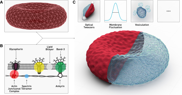
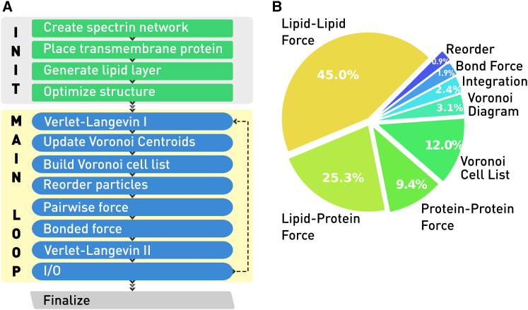
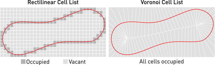
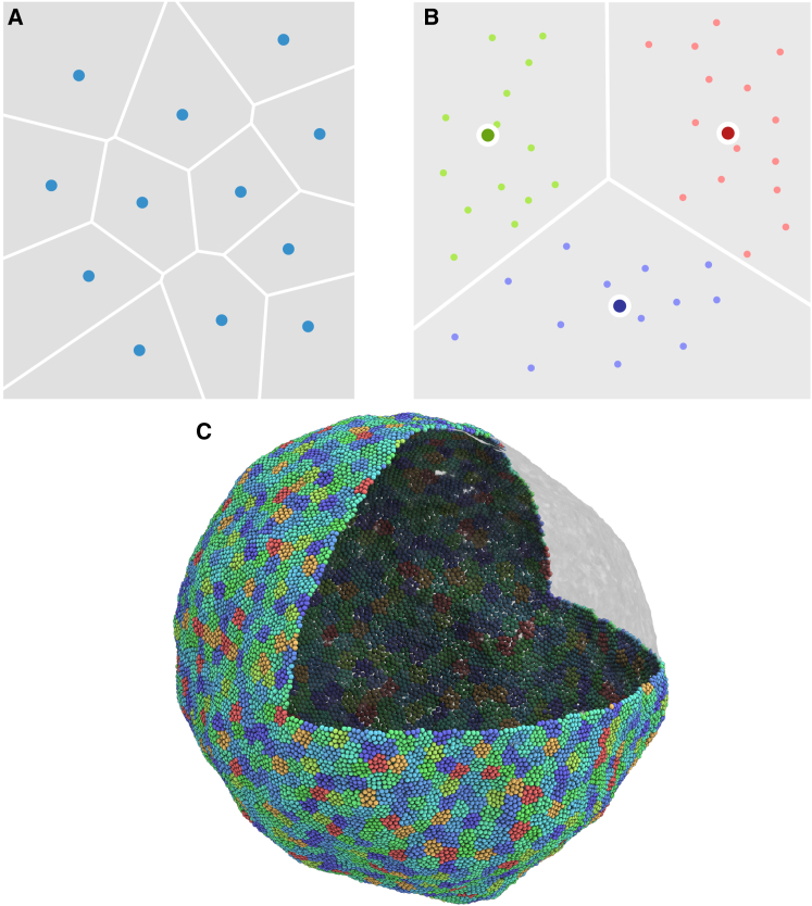
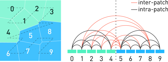
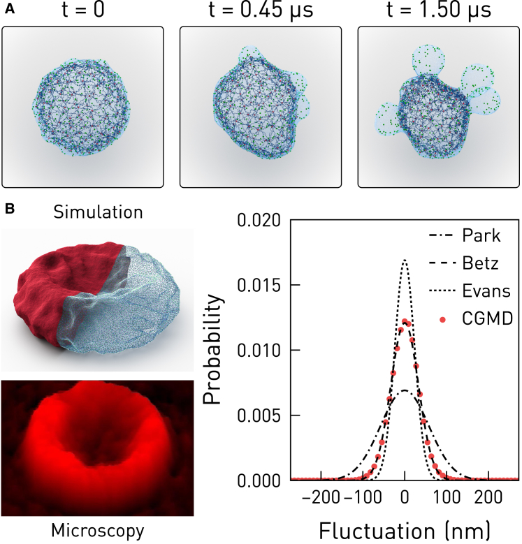
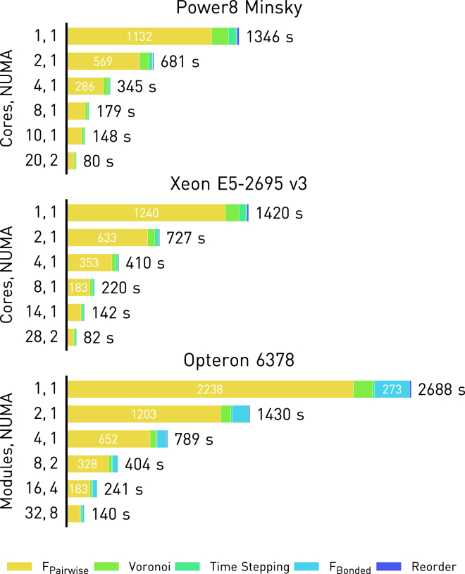
We present OpenRBC, a coarse-grained molecular dynamics code, which is capable of performing an unprecedented in silico experiment-simulating an entire mammal red blood cell lipid bilayer and cytoskeleton as modeled by multiple millions of mesoscopic particles-using a single shared memory commodity workstation. To achieve this, we invented an adaptive spatial-searching algorithm to accelerate the computation of short-range pairwise interactions in an extremely sparse three-dimensional space. The algorithm is based on a Voronoi partitioning of the point cloud of coarse-grained particles, and is continuously updated over the course of the simulation. The algorithm enables the construction of the key spatial searching data structure in our code, i.e., a lattice-free cell list, with a time and space cost linearly proportional to the number of particles in the system. The position and the shape of the cells also adapt automatically to the local density and curvature. The code implements OpenMP parallelization and scales to hundreds of hardware threads. It outperforms a legacy simulator by almost an order of magnitude in time-to-solution and >40 times in problem size, thus providing, to our knowledge, a new platform for probing the biomechanics of red blood cells.
Copyright © 2017 Biophysical Society. Published by Elsevier Inc. All rights reserved.
Figures
References
-
- Feng F., Klug W.S. Finite element modeling of lipid bilayer membranes. J. Comput. Phys. 2006;220:394–408.
-
- Powers T.R., Huber G., Goldstein R.E. Fluid-membrane tethers: minimal surfaces and elastic boundary layers. Phys. Rev. E Stat. Nonlin. Soft Matter Phys. 2002;65:041901. - PubMed
-
- Helfrich W. Elastic properties of lipid bilayers: theory and possible experiments. Z. Naturforsch. C. 1973;28:693–703. - PubMed
-
- Feller S.E. Molecular dynamics simulations of lipid bilayers. Curr. Opin. Colloid Interface Sci. 2000;5:217–223.
Publication types
MeSH terms
Grants and funding
LinkOut - more resources
Full Text Sources
Other Literature Sources
