

The COMPLEXity in herpesvirus entry
- PMID: 28538165
- PMCID: PMC8601108
- DOI: 10.1016/j.coviro.2017.04.006
The COMPLEXity in herpesvirus entry
Abstract
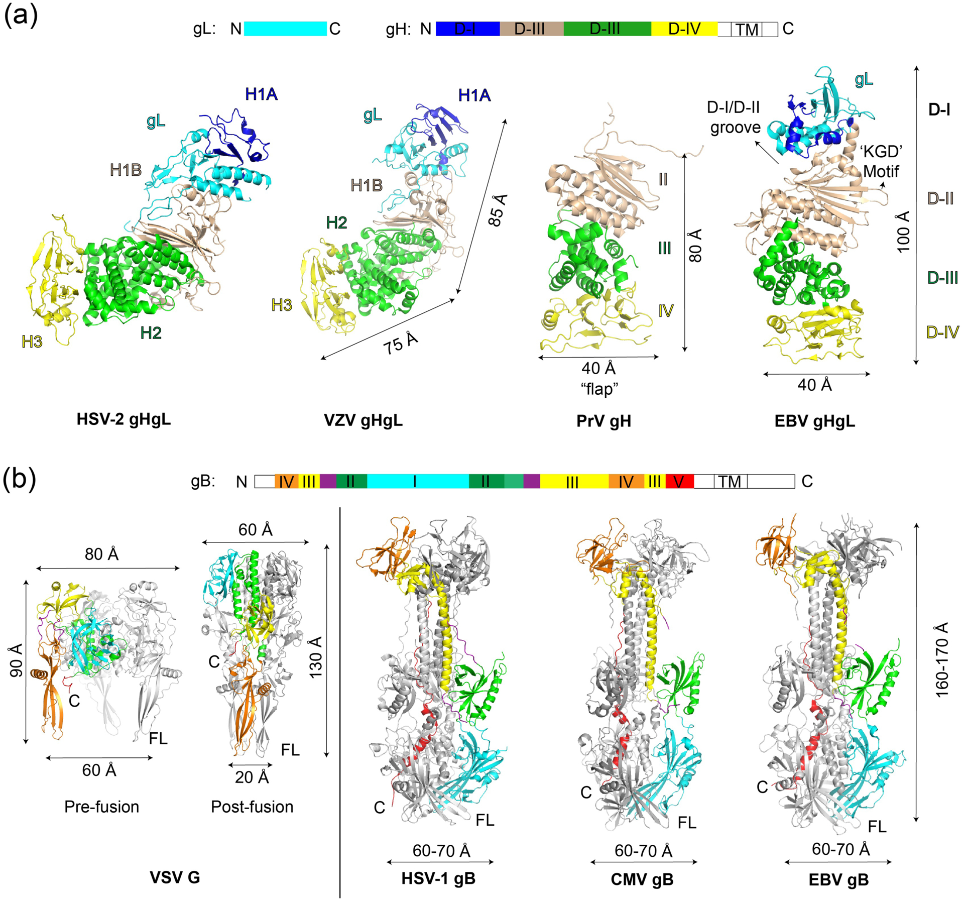
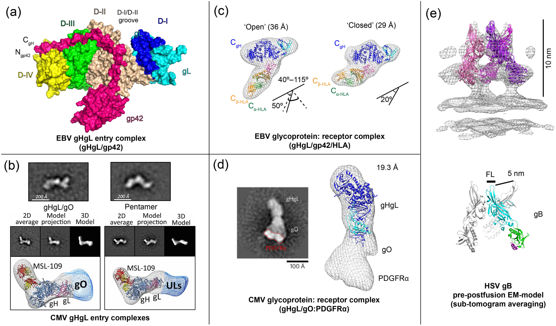
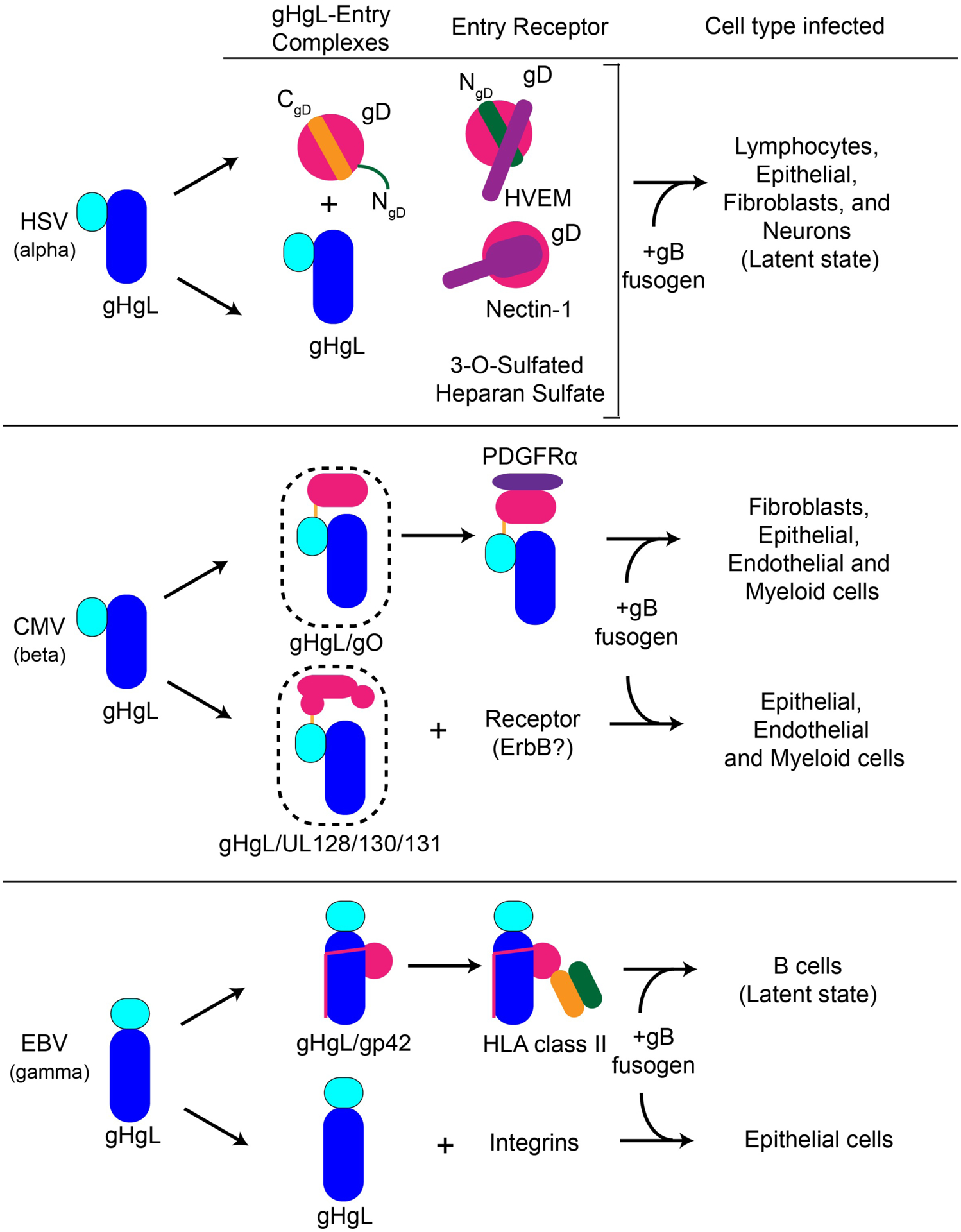
Enveloped viruses have evolved diverse transmembrane proteins and protein complexes to enable host cell entry by regulating and activating membrane fusion in a target cell-specific manner. In general terms, the entry process requires a receptor binding step, an activation step and a membrane fusion step, which can be encoded within a single viral protein or distributed among multiple viral proteins. HIV and influenza virus, for example, encode all of these functions in a single trimeric glycoprotein, HIV env or influenza virus hemagglutinin (HA). In contrast, herpesviruses have the host receptor binding, activation and fusogenic roles distributed among multiple envelope glycoproteins (ranging from three to six), which must coordinate their functions at the site of fusion. Despite the apparent complexity in the number of viral entry proteins, herpesvirus entry is fundamentally built around two core glycoprotein entities: the gHgL complex, which appears to act as an 'activator' of entry, and the gB protein, which is thought to act as the membrane 'fusogen'. Both are required for all herpesvirus fusion and entry. In many herpesviruses, gHgL either binds host receptors directly or assembles into larger complexes with additional viral proteins that bind host receptors, conferring specificity to the cells that are targeted for infection. These gHgL entry complexes (ECs) are centrally important to activating gB-mediated membrane fusion and establishing viral tropism, forming membrane bridging intermediates before gB triggering. Here we review recent structural and functional studies of Epstein-Barr virus (EBV) and Cytomegalovirus (CMV) gHgL complexes that provide a framework for understanding the role of gHgL in herpesvirus entry. Furthermore, a recently determined EM model of Herpes Simplex virus (HSV) gB embedded in exosomes highlights how gB conformational changes may promote viral and cellular membrane fusion.
Copyright © 2017. Published by Elsevier B.V.
Conflict of interest statement
Figures
References
-
- Chesnokova LS, Jiang R, Hutt-Fletcher LM: Viral Entry. Curr. Top. Microbiol. Immunol 2015, 391:221–35. - PubMed
Publication types
MeSH terms
Substances
Grants and funding
LinkOut - more resources
Full Text Sources
Other Literature Sources
