. 2017 May 23;19(8):1723-1738.
doi: 10.1016/j.celrep.2017.05.006.

Identification and Characterization of a Class of MALAT1-like Genomic Loci

Affiliations

Identification and Characterization of a Class of MALAT1-like Genomic Loci

Bin Zhang et al. Cell Rep. .

Abstract

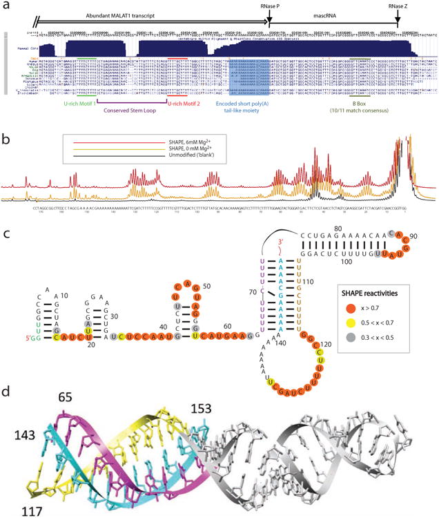
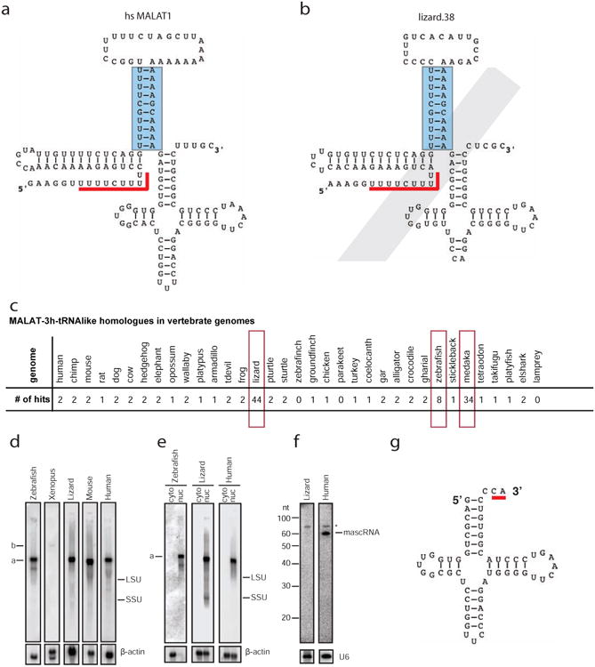
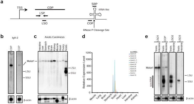
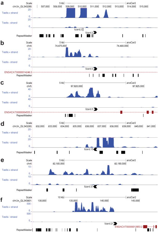
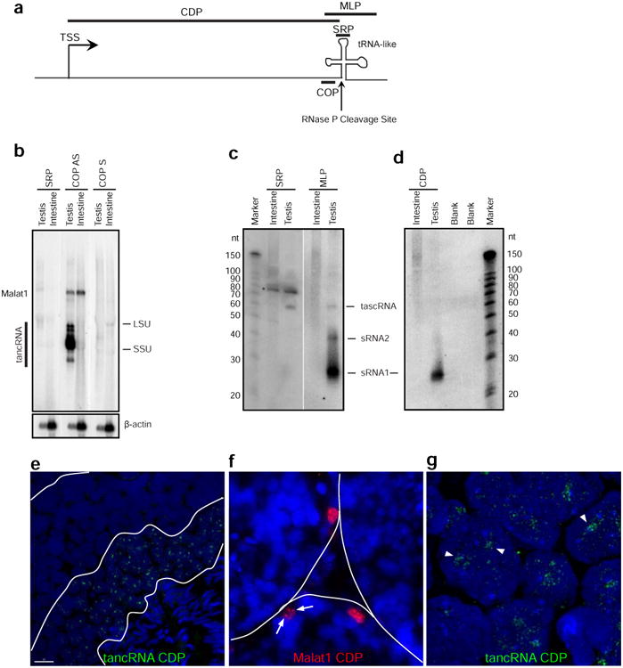
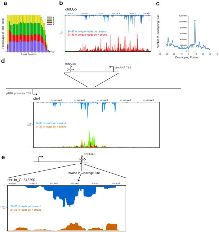
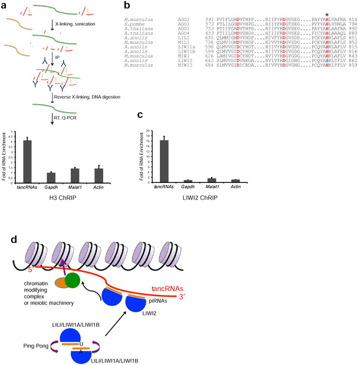
The MALAT1 (Metastasis-Associated Lung Adenocarcinoma Transcript 1) gene encodes a noncoding RNA that is processed into a long nuclear retained transcript (MALAT1) and a small cytoplasmic tRNA-like transcript (mascRNA). Using an RNA sequence- and structure-based covariance model, we identified more than 130 genomic loci in vertebrate genomes containing the MALAT1 3' end triple-helix structure and its immediate downstream tRNA-like structure, including 44 in the green lizard Anolis carolinensis. Structural and computational analyses revealed a co-occurrence of components of the 3' end module. MALAT1-like genes in Anolis carolinensis are highly expressed in adult testis, thus we named them testis-abundant long noncoding RNAs (tancRNAs). MALAT1-like loci also produce multiple small RNA species, including PIWI-interacting RNAs (piRNAs), from the antisense strand. The 3' ends of tancRNAs serve as potential targets for the PIWI-piRNA complex. Thus, we have identified an evolutionarily conserved class of long noncoding RNAs (lncRNAs) with similar structural constraints, post-transcriptional processing, and subcellular localization and a distinct function in spermatocytes.

Keywords: Anolis carolinesis; MALAT1; PIWI; evolution; lizard; long noncoding RNA; piRNA; tRNA-like; testis; triple helix.


Figures

Figure 1
Figure 1. The 3′ end of MALAT1 forms a triple helix structure
(a) The sequence conservation of the 3′ end of MALAT1. (b) SHAPE chemical probing of the 3′ end of human MALAT1 is consistent with a triple helix. Capillary electrophoresis traces display nucleotides with low and high SHAPE reactivity, corresponding to low and high flexibility. Black, unmodified control; gold, SHAPE reactivity for 0 mM Mg2+; red, SHAPE reactivity for 6 mM Mg2+. (c) Secondary structure of the 3′ end of human MALAT1, as inferred by SHAPE and DMS probing experiments (see supplementary data for DMS). Orange, high reactivity; yellow, medium reactivity; grey, low reactivity; un-circled nucleotides, near-zero reactivity. (d) Tertiary structure model of the 3′ end of human MALAT1 demonstrates stereochemical feasibility of a triple helix. Purple, yellow and cyan strand correspond to similarly colored strands in (c).
Figure 2
Figure 2. A class of genomic loci with structures similar to the MALAT1 3′ end was identified in vertebrate genomes
(a) The primary sequence and predicted secondary structure of human MALAT1 3′ module as identified by a homology search using Infernal 1.1rc1 against the human genome using CM file MALAT1-3h-tRNAlike.cm (Supplementary Data SF4). Only basepairs modeled by the CM are indicated with lines connecting two nucleotides. The triple helix is formed between the stem structure indicated by a blue rectangle and its upstream U-rich motif indicated by a red line. (b) One MALAT1 3′ end homologue (lizard.38) with a degenerative tRNA-like structure that was identified in the lizard genome using the MALAT1-3h-tRNAlike.cm CM profile. (c) Summary of MALAT1-3h-tRNAlike hits with an E value less than 1×10-11 in 35 genomes (see Supplementary Data SF7 and SF12). Note that most mammals have one or two hits, while lizard, zebrafish, and medaka have more than two MALAT1-3h-tRNAlike hits (highlighted in red open rectangles). (d) Northern blot analysis shows that MALAT1 orthologues were expressed from cultured cells of zebrafish (ZFL), Xenopus (A6), lizard (IgH-2), mouse (C2C12) and human (U2OS). (e) Northern blot analysis shows that MALAT1 is enriched in nuclei of cultured cells of zebrafish (ZFL), lizard (IgH-2), and human (U2OS). (f) Small RNA Northern blot analysis shows that lizard mascRNA is not detectable in the IgH-2 cell line. (g) Secondary structure of lizard mascRNA in the IgH-2 cell line with noncanonical CA tail modification indicated by a red line. LSU, large subunit of ribosomal RNA; SSU, small subunit of ribosomal RNA; cyto, cytoplasmic fraction; nuc, nuclear fraction; *, non-specific band. β-actin and U6 are loading controls.
Figure 3
Figure 3. Long noncoding RNAs are produced from MALAT1-like genomic loci in Anolis carolinesis in a tissue-specific manner
(a) Schematic of the transcriptional unit of the Malat1 3′ module homologous loci. TSS, transcriptional start site; CDP, IgH-2 cDNA Probe (used in panel b); COP, Consensus Oligonucleotide Probe that is derived from the multi-sequence alignment of 44 homologues (used in panels b, c, and e); LSP, locus-specific primers (used in panel d); LSO, locus-specific oligonucleotide (used in panel e); SRP, small RNA probe; DSO, downstream oligonucleotide; the vertical arrow, the predicted RNase P cleavage site just before the tRNA-like structure. (b) Northern blot analysis shows that MALAT1 RNA is highly expressed in lizard IgH2 cells detected by both cDNA probe and COP, but no other RNA species are detected by the COP. (c) Northern blot analysis shows that COP labels MALAT1 in all tissues of Anolis carolinensis and multiple RNA species in testis (named as tancRNAs). (d) qRT-PCR analysis using locus-specific primer (LSP) sets shows that tancRNAs from different genomic loci are specifically expressed in lizard testis. Y axis, relative expression level; X axis, different tissues. (e) Northern blot analysis shows that tancRNAs are expressed from different genomic loci. Note that LSO1 recognizes MALAT1, LSO2 recognizes multiple tancRNA species (lizard.20, lizard. 12, lizard.8), and LSO3 detects one major tancRNA species (lizard.30). β-actin is the loading control.
Figure 4
Figure 4. Different examples of transcripts from MALAT1-like genomic loci
(a) – (f) Normalized RNA-Seq reads from testis (upper track: reads on sense strand, lower track: reads on antisense strand). Black arrowheads denote MALAT1-3h-tRNAlike loci and their direction of transcription. Chromosome location and annotated Ensembl transcripts are indicated. Read depth scale is displayed on the y-axis. Interspersed repeats and low complexity DNA Sequences are indicated in the RepatMasker track. 6 examples are shown; nomenclature according to Supplementary Data SF7.
Figure 5
Figure 5. tancRNAs are processed and enriched in nuclei and multiple small RNA species are produced from tancRNA loci
(a) Schematic of the transcriptional unit at the Malat1 3′ module homologous loci. TSS, transcriptional start site; CDP, cDNA Probe (used in panels d, e, f, and g); MLP, MALAT1 3′-like probe (used in panel c); the vertical arrow, the predicted RNase P cleavage site just before the tRNA-like structure. (b) tancRNAs were processed as human MALAT1. Large RNA Northern blot analysis shows that SRP (antisense of mascRNA/tascRNA) does not recognize any significant bands (lane 1). COP AS (tancRNA consensus antisense probe) detects both MALAT1 and tancRNAs (lane 2), while COP S (tancRNA consensus sense probe) does not recognize any significant bands (lane 3). (c) Multiple small RNA species were produced from tancRNA loci. Small RNA Northern blot analysis shows that SRP (mascRNA/tascRNA antisense oligonucleotide probe) detects mascRNA/tascRNA in lizard testis (left), while MLP (consensus PCR pooled probe) detects three small RNA species with sizes of 26, 40, and 60 nt (right). (d) Small RNAs with the size of piRNAs were produced from tancRNA loci. Small RNA Northern blot analysis detects a strong RNA band with the size of 26 nt using CDP (a cDNA probe from the lizard.11 locus, see Supplementary Data SF7 and SF12). (e) RNA FISH shows that tancRNAs are localized to nuclei of pachytene and/or round spermatocytes in adult lizard testis. (f) RNA FISH shows that MALAT1 is enriched in nuclei of the periphery of seminiferous tubules. Arrows indicate nuclear punctuate signal pattern. (g) High-resolution imaging of RNA FISH (in e) shows that tancRNAs occupies distinctive nuclear domains (arrowheads). LSU, large subunit of ribosomal RNA; SSU, small subunit of ribosomal RNA. Equal amount of total RNAs were loaded for each lane. β-actin is the loading control. *, non-specific band.
Figure 6
Figure 6. piRNAs are produced from the opposite strand of tancRNAs
(a) Positional distribution of nucleotide composition of small RNAs sequenced from adult lizard testis. A strong 1U bias and a weak 10A enrichment were noticed. (b) Small RNAs with the size of 24-33 nt were uniquely mapped to a dual-strand piRNA cluster on chrLGb. (c) A strong ping-pong signature of piRNAs from the dual-strand cluster on chrLGb (see b). X axis, overlapping index; Y axis, number of overlapping pairs. (d) piRNAs mapped to a tancRNA locus (lizard.37, see Supplementary Data SF7 and SF12) on chr4. Orange, unique mapper on – strand; blue, multiple mapper on – strand; red, unique mapper on + strand; green, multiple mapper on + strand. (e) piRNAs mapped to the 3′ end of a tancRNA locus (lizard.2, see Supplemental File SF7 and SF12) on chrUn_GL343290 with multiple mappers (reporting = 50).
Figure 7
Figure 7. 3′-end of tancRNAs act as targets of antisense piRNAs
(a) A schematic diagram of Chromatin RNA Immunoprecipitation (ChRIP) analysis. ChRIP using antibody against Histone H3 shows that tancRNAs (primer sequences in Supplementary Data SF14) is associated with chromatin. (b) Sequence alignment showing the catalytic triad (DDH motif) of known Slicers and MIWI. LIWI2 has a variant of the third His to Ser (H856S; marked with an asterisk). (c) ChRIP analysis using antibody against LIWI2 shows that tancRNAs is associated with LIWI2. X axis, RNA examined; Y axis, Fold of RNA enrichment. Sample size n = 3. (d) A proposed working model of tancRNA-piRNA-Liwi2 complex. TancRNAs act as targets of PIWI-piRNA complexes through their 3′ end triplex structure. 5′ end tancRNAs serve as domains to interact with DNA and/or chromatin, therefore tethering PIWI-piRNA complexes to specific genomic loci to regulate chromatin activity.

Similar articles

Cited by

References

    1. Aravin A, Gaidatzis D, Pfeffer S, Lagos-Quintana M, Landgraf P, Iovino N, Morris P, Brownstein MJ, Kuramochi-Miyagawa S, Nakano T, et al. A novel class of small RNAs bind to MILI protein in mouse testes. Nature. 2006;442:203–207. - PubMed
    1. Aravin AA, Hannon GJ. Small RNA silencing pathways in germ and stem cells. Cold Spring Harb Symp Quant Biol. 2008;73:283–290. - PubMed
    1. Armour CD, Castle JC, Chen R, Babak T, Loerch P, Jackson S, Shah JK, Dey J, Rohl CA, Johnson JM, et al. Digital transcriptome profiling using selective hexamer priming for cDNA synthesis. Nature methods. 2009;6:647–649. - PubMed
    1. Arun G, Diermeier S, Akerman M, Chang KC, Wilkinson JE, Hearn S, Kim Y, MacLeod AR, Krainer AR, Norton L, et al. Differentiation of mammary tumors and reduction in metastasis upon Malat1 lncRNA loss. Genes Dev. 2016;30:34–51. - PMC - PubMed
    1. Batista PJ, Chang HY. Long noncoding RNAs: cellular address codes in development and disease. Cell. 2013;152:1298–1307. - PMC - PubMed

Publication types