

Magnetic resonance imaging evaluation of the labrum to predict acetabular development in developmental dysplasia of the hip: A STROBE compliant study
- PMID: 28538419
- PMCID: PMC5457899
- DOI: 10.1097/MD.0000000000007013
Magnetic resonance imaging evaluation of the labrum to predict acetabular development in developmental dysplasia of the hip: A STROBE compliant study
Abstract
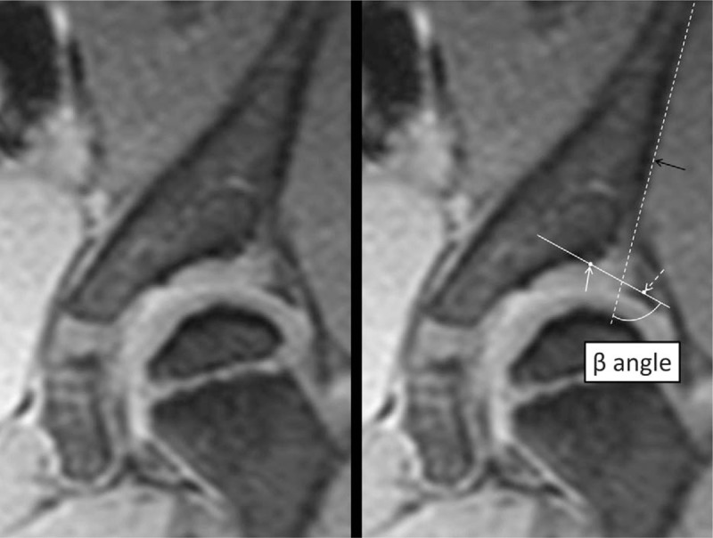
Recently, more attention has been paid to the role of the acetabular labrum. Therefore, we designed a retrospective cohort study of patients with residual hip dysplasia (RHD) who underwent magnetic resonance imaging (MRI). The objective of this study was to investigate an association between the MRI appearance of the labrum before school age and the natural history of RHD.We retrospectively investigated 45 hips of 40 patients who underwent MRI at about 3 and 4 years of age for RHD and were conservatively followed up with until 6 years of age or older. We evaluated the extent of eversion with a new method that measures the β angle (MRI β angle) using landmarks of the Graf method on MRI T2*-weighted images. The outcome measure was the Severin classification at the final follow-up. We compared the radiographic and MRI parameters at approximately 3 and 4 years of age between the good and poor outcome groups. The Student t test or one-way analysis of variance was used to compare the quantitative variables between groups. The Chi-square test was used to perform a ratio comparison.Although there was a significant difference in the center-edge (CE) angle, there was no significant difference in the acetabular index and the ratio of the presence of femoral head necrosis and the break in Shenton line between the good and poor groups. The MRI β angle was significantly greater in the poor outcome group than in the normal and good outcome groups. The cut-off value of the MRI β angle to differentiate the good and poor outcome groups was 65°, and its specificity and sensitivity were 92% and 53%, respectively.There was labral eversion on MRI scans in patients with RHD. Acetabular development before adolescence was poorer with greater labral eversion on MRI scans. The specificity for poor acetabular development was high when the MRI β angle was 65° or more. The MRI β angle has the potential to predict acetabular development.
Conflict of interest statement
The authors have no conflicts of interest to disclose.
Figures


References
-
- Salter RB. The classic. Innominate osteotomy in the treatment of congenital dislocation and subluxation of the hip. J Bone Joint Surg Br 1961;43:518–39.
-
- Wiberg G. Studies on dysplastic acetabula and congenital subluxation of the hip joint. Acta Chir Scand 1939;83:1.
-
- Albinana J, Dolan LA, Spratt KF, et al. Acetabular dysplasia after treatment for developmental dysplasia of the hip. Implications for secondary procedures. J Bone Joint Surg Br 2004;86:876–86. - PubMed
-
- Hattori T, Ono Y, Kitakoji T, et al. Soft-tissue interposition after closed reduction in developmental dysplasia of the hip. The long-term effect on acetabular development and avascular necrosis. J Bone Joint Surg Br 1999;81:385–91. - PubMed
-
- Kitoh H, Kitakoji T, Katoh M, et al. Prediction of acetabular development after closed reduction by overhead traction in developmental dysplasia of the hip. J Orthop Sci 2006;11:473–7. - PubMed
Publication types
MeSH terms
LinkOut - more resources
Full Text Sources
Other Literature Sources
Medical
